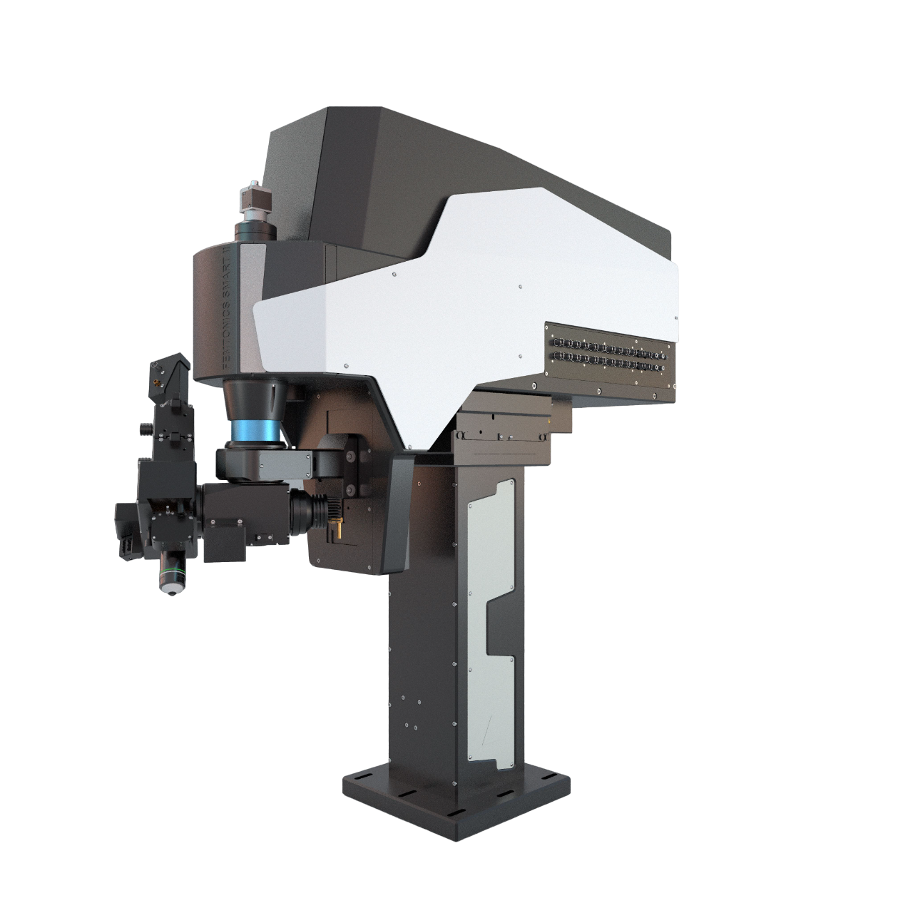
3P imaging
Three-photon (3P) microscopy allows noninvasive structural and functional imaging by making cells visible in deep tissues with high spatial resolution and better contrast compared to two-photon excitation. The optical design required for 3P excitation establishes higher axial resolution than that of two-photon microscopy. The spectral window enables three-photon excitation of a variety of fluorophores, such as the current generations of protein-based genetically encoded calcium indicators (e.g. GCaMP6) and the repetition rate of the laser source is adequate for imaging Ca2+ transients produced from neural activity.
The video shows an approximately 1000 μm thick 3P z-stack from a mouse cortex. Purple: blood vessels, green: GFP labelled pericytes. Footage courtesy of Dr. Severin Filser.

3P range microscopy
2D Multiple-Line Scanning by FEMTOSmart GALVO
The adaptable X and Y mirrors of the galvanometric scanner, coupled with the special electronic boards from Femtonics, can follow the tortuous protrusions of the dendritic arbor precisely using the 2D random-access point, multiple-line and folded frame scanning methods. By limiting the scanning to the interesting dendritic segments carrying spines and omitting the space between them, both the scanning speed (up to 2 kHz) and the SNR can be increased using 2D random-access point and multiple-line scanning modes. The advantage is multiple-line scanning is the imaging without interruption at multiple dendritic branches, it is the cost-effective, alternative solution compared to the acousto-optic scanner-based line scanning methods.
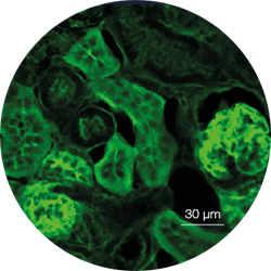
Third Harmonic Generation
Third Harmonic Generation is a specific effect of 3P excitation, that results from the conversion of three incoming photons into one emitted photon with tripled energy and thus, the emission of the light of one-third the wavelength. THG occurs at structural interfaces that are formed between aqueous fluids and lipid-rich structures, for instance, biological membranes, and between water and large protein aggregates such as collagen bundles or muscle fibers.
The figure shows a THG image of a section of a mouse kidney: kidney cells were excited at 1500 nm, and the emitted photons were collected at the green channel (~500 nm) of the detector system.
“We have found a massively great partner in Femtonics and acquiring the groundbreaking 3P (three-photon) FEMTO SMART system ensures never-seen experimental results in deep imaging. We are impressed how fast they are reacting at our requests and able to think together with us on scientific solutions. Their technical level and support are professional, and it is always good to have a trustful partner in daily scientific life.”
Dr. Severin Filser
DZNE – German Center for Neurodegenerative Diseases, Germany