Deep functional imaging
One of the most significant advantages of two-photon microscopy is the ability to examine not only the tissue’s surface but also its deeper layers. While confocal microscopy can penetrate the tissue, it is limited to just a few dozen micrometers.
Thus, a fundamental question arises: How deep can we effectively visualize within the brain?
The answer depends on a multitude of external parameters, including the laser’s wavelength and type, quality of labeling, tissue type, and preparation quality, among others. Furthermore, the imaging solutions themselves play a pivotal role in achieving depths surpassing even 1mm from the surface. In this context, we present our approach to Deep Functional Imaging.
Deeper cortical layers and the hippocampus are widely investigated brain regions. Deep Functional Imaging with the FEMTO3D Atlas and FEMTO3D Atlas Plug & Play
provides fast, 3D imaging means for different deeper brain regions as well.
Several technical elements contribute to the enhanced depth penetration capability of the microscope, all of which are integrated in FEMTO3D Atlas and FEMTO3D Atlas Plug & Play, along with their accompanying driving software, MES:
• Proper dispersion compensation
• Lateral and depth-dependent intensity compensation
• Large NA, non-descanned detectors optimized for scattered photons
• High-quality optimized filter sets
• High QE GaAsP detector modules
• Depth-dependent beam expansion
When reaching deeper regions, photon scattering becomes more prominent at the edge of the beam cone, particularly with high Numerical Aperture (NA) objectives. Consequently, imaging intensity substantially decreases. To address this issue, it is crucial to reduce the diameter of the imaging laser beam. A narrower laser beam minimizes scattering, thus enhancing image quality at greater tissue depths (Figure 1).
Features:
- Fully integrated into 4D BCU and MES.
- Convenient, direct control of beam diameter at the objective pupil.
- The low pointing provided by the sliding lens mechanism is fully compensated by the beam stabilization system of the Atlas scanhead.
- Wavelength-independent beam diameter adjustment.
- Calibrated for the specific laser.
- Optimized and AR coated for the 750-1050 nm wavelength range.
- Minimal focus shift under the objective, easily compensated by objective movement or AO focusing.
- Programmatically adjustable during z-stacks and metaprotocols.
- Can be installed as an upgrade – contact us for details.
Benefits:
- Precise control of effective excitation numerical aperture.
- Penetration depth of up to 1 mm.
- Reduced power losses due to scattering during deep imaging.
- Fine-tuning of focal spot (PSF) size to compensate for motion artifacts or increase the volume of imaging or photostimulation.
- Decreased power losses from aberrations (resulting from uncorrected cover glass).
- Enhanced transmission for medium and high magnification objectives.
- Expanded effective acousto-optic Z (remote) focusing range attributed to improved transmission, even for higher |Z| values.
Depth over everything
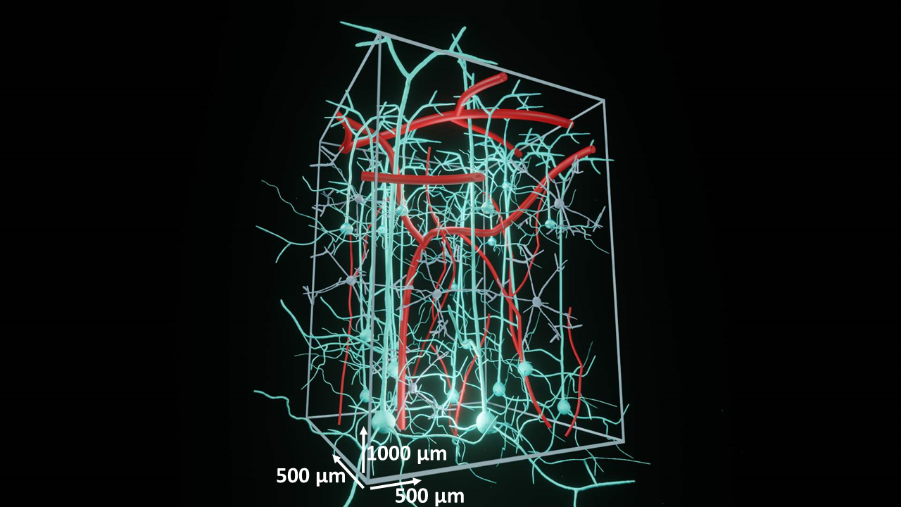
Through the application of precise staining techniques, Deep Functional Imaging (DFI) offers the potential to delve into tissue structures up to their physical limits. The wavelength of light plays a significant role in the depth of imaging, with longer wavelengths allowing for deeper penetration. When measured from the surface, it becomes feasible to achieve a depth of 1 mm with an optimized beam diameter (Figure 2). This capability extends to visualizing brain mechanisms during in vivo awake experiments.
Greater intensity directly corresponds to increased information acquisition during imaging. Hippocampal imaging serves as an excellent example to underscore the potential of DFI (Figure 2). By avoiding tissue damage, a higher Signal-to-Noise Ratio (SNR) can be attained, enabling longer acquisition times and enhanced spatial resolution.
Examples of Deep Functional Imaging in the mouse neocortex