Blood flow tracking
Following blood flow deep in the living tissue
Abnormalities in hemodynamics are linked to various disease states, such as stroke, Alzheimer’s, etc., however, to better understand these associations, more precise optical techniques are needed for interrogating blood flow velocity in vivo in 3D. Acousto-optic (AO) multiphoton microscopy is a superior technique to perform blood flow tracking at the cellular level, at a high spatiotemporal resolution deep in the living 3D tissue. The unique scanning modes of the FEMTO3D Atlas allow you to record continuously along intricate 3D patterns, and its extensions make it possible to follow multiple parameters at the same time, like blood flow and cellular activity. These applications can be useful in various fields, such as neuroscience, immunology, dermatology, oncology and more.
FEMTO3D Atlas Dichro with two laser sources for dual excitation
This extension of the FEMTO3D Atlas platform enables fast 3D region of interest (ROI) scanning in a large 3D volume, as well as a quasi-simultaneous detection of neuronal activity and blood flow using two different laser sources. The Femtonics software package provides built-in ROI and curve analysis options, meeting all needs.
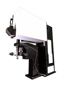
Microscope equipment
- FEMTO3D Atlas acousto-optic microscope capable of rapid XY and Z focusing (3D imaging)
- 4D Beam Conditioning Unit for the stabilization of the laser beams
- Two femtosecond laser sources of different wavelengths
- Dichro extension for fast switching between the two lasers line for quasi-simultaneous dual excitation
Technical procedure for acquisition and analysis
Here we injected a red dextran intravenously to make blood vessels appear in red. The passing red blood cells (RBCs) show as shadows in the red stream (Fig1.A). This way, the speed of the blood stream can be measured by following an RBC in a time-dependent manner (Fig 1.B).
Rapid ROI imaging by the state-of the-art FEMTO3D Atlas AO platform enables 3D ribbon scanning, following vessels through multiple cortical layers in a large brain volume. The total length of the ribbons can reach up to ~5 mm, while maintaining a resolution and speed sufficient to measure the velocity of blood flow (Fig 2.)
Simultaneous measurement of neuronal activity and blood flow velocity
Research of cardiac diseases may be supported by a combined method of simultaneous Ca2+ activity and blood flow imaging. This provides a novel opportunity of acquiring local information about neuronal and cardiovascular status. Using the FEMTO3D Atlas Dichro with two laser-sources of different wavelength, blood cell movement and neuronal activity can be measured at the same time. Fast switching between the two laser sources makes it possible to image quasi-simultaneously on two different emission wavelengths. This method can be useful for studying the detrimental consequences of stroke on vascular condition, neuronal damage, and even regeneration processes (Fig 3.).