Dendritic Imaging
Dendrites and dendritic spines are made of fine, thin and elongated segments therefore they are difficult to study. However, using two-photon technology, we are able to collect signals from deeper regions of the brain, while avoiding phototoxicity. Additionally, spatially confined, regional scanning of axons, dendrites and spines allows detecting even subthreshold signals with high signal-to-noise ratio (SNR). We have several configurations aiding to visualize dendritic arborization, and perform functional measurements in vivo and in vitro. Now, with the help of our new real time motion-correction called Femtonics 3D Real-Time Motion Correction motion artifacts even in vivo in dendritic measurements will no longer be a problem!
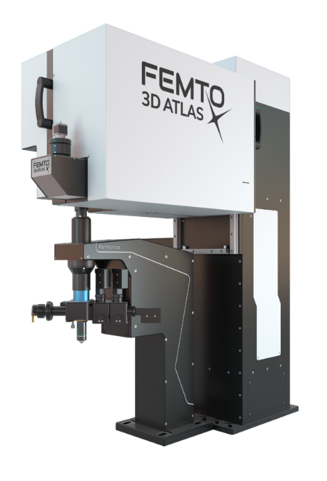
With the FEMTO3D Atlas point scanning speeds up to a 100kHz can be achieved, which helps determining activity at virtually any sites on dendrites. 3D random-access point scanning and its advanced versions support all kinds of functional dendritic imaging from single spine investigations to full arborization measurements, even combined with photostimulation solutions.
Fast drift along the dendrite
3D trajectory scanning
Dendritic and spine activity during motions
3D ribbon scanning
An extension of the 3D multiple-line scanning is 3D ribbon scanning, which makes it possible to image ribbon shaped surfaces containing dendrites and the neighboring areas. Figures show 3D ribbons encompassing seven dendritic segments with their spines of a GCaMP6-labeled layer II/III pyramidal neuron measured within the brain of a living mouse.
Real-time motion-correction with the Femtonics 3D Real-Time Motion Correction
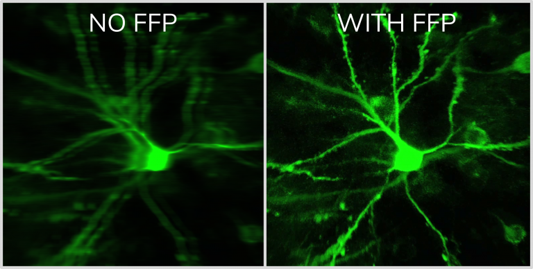
Our solution takes advantage of acousto-optics technology and elevates it to a whole new level. The flexible drift scanning method creates the opportunity to perform correction calculations with a predefined and adjustable frequency. The algorithm is based on correction calculations derived from selecting stable objects within the field of view (FOV), which serve as reference points during scanning. With our method providing an effortless means to scan in any direction in 3D, reference measurements are equally efficient in the Z direction as they are in the XY direction, which is a unique feature of the Femtonics 3D Real-Time Motion Correction.
High-speed arbitrary frame scanning
High-speed arbitrary frame scanning gives you the freedom to tilt and rotate the scanning plane in any direction. With this feature, in vivo imaging of long dendrites can be performed in a single plane. To examine multiple neurons’ apical dendrites simultaneously, the tilted frame can be extended into a volume element.
The video shows the apical dendrites of the neurons getting aligned with the scanning plane, which means the spatio-temporal activity patterns of these long segments passing multiple cortical layers can be recorded without requiring lengthy and painstaking optical sectioning.
FemtoSmart offers numerous imaging opportunities and thanks to its modularity researchers can study dendrites with spines from wide variations of perspectives.
The fastest in 2D
2D Multiple-Line Scanning by FEMTOSmart GALVO
The adaptable X and Y mirrors of the galvanometric scanner, coupled with the special electronic boards from Femtonics, can follow the tortuous protrusions of the dendritic arbor precisely using the 2D random-access point, multiple-line and folded frame scanning methods. By limiting the scanning to the interesting dendritic segments carrying spines and omitting the space between them, both the scanning speed (up to 2 kHz) and the SNR can be increased using 2D random-access point and multiple-line scanning modes. The advantage is multiple-line scanning is the imaging without interruption at multiple dendritic branches, it is the cost-effective, alternative solution compared to the acousto-optic scanner-based line scanning methods.
Imaging dendrites and spines with motion correction
Folded frame scanning by FEMTOSmart GALVO
Using folded frame scanning, an area along a pre-selected line can be imaged. The selected regions can take many shapes, from areas around straight lines to complex bent curves. This advanced scanning method is useful for following events along curved dendrites with spines, and can also be advantageous for dendritic measurements in behaving animal models where motion artefacts are a common problem. The images are corrected for motion offline by the control software, as long as the dendrite remains in the scanned area.
Coming Soon: Automated dendrite selection tool
As dendritic segments are abundant, complex, 3 dimensional features of the brain, selecting ROIs around them is a maticulous job, reliant on human perception. With the help of this optional module, you will be able select all dendritic segments in your field of view, just with some push of buttons. This will save you time and energy, and the end result will be more precisely aligned, down to the pixel level!
References
Pyramidal neurons form active, transient, multilayered circuits perturbed by autism-associated mutations at the inception of neocortex Munz M, Bharioke A, Kosche G, Moreno-Juan V, Brignall A, Rodrigues TM, Graff-Meyer A, Ulmer T, Haeuselmann S, Pavlinic D, Ledergerber N, Gross-Scherf B, Rózsa B, Krol J, Picelli S, Cowan CS, Roska B
Fast 3D Imaging of Spine, Dendritic, and Neuronal Assemblies in Behaving Animals. Gergely Szalay, Linda Judak, Gergely Katona, Katalin Ocsai, Gabor Juhasz, Mate Veress, Zoltan Szadai, Andras Feher, Tamas Tompa, Balazs Chiovini, Pal Maak, Balazs Rozsa, Neuron (2016)
Dendritic spikes induce ripples in parvalbumin interneurons during hippocampal sharp waves. B Chiovini, G F Turi, G Katona, A Kaszas, D Palfi, P Maak, G Szalay, M F Szabo, Z Szadai, Sz Kali and B Rozsa, Neuron (2014)
Fast two-photon in vivo imaging with three-dimensional random-access scanning in large tissue volumes. Gergely Katona, Gergely Szalay, Pál Maak, Attila Kaszas, Mate Veress, Daniel Hillier, Balazs Chiovini, E Sylvester Vizi, Botond Roska & Balazs Rozsa, Nature Methods (2012)
Matching Cell Type to Function in Cortical Circuits. Luc Estebanez, Jens Kremkow, James F.A. Poulet, Neuron (2015)
In Vivo Monosynaptic Excitatory Transmission between Layer 2 Cortical Pyramidal Neurons. Jean-Sebastien Jouhanneau, Jens Kremkow, Anja L. Dorrn, James F.A. Poulet, Cell Reports (2015)
Roller Coaster Scanning reveals spontaneous triggering of dendritic spikes in CA1 interneurons. G. Katona, A. Kaszas, G. F. Turi, N. Hajos, G. Tamas, E.S. Vizi, B. Rozsa, PNAS (2011)