Award winning 3D real-time motion correction
Femtonics has won the Innovation Award at the 32nd Hungarian Innovation Grand Award competition with the “Femtonics FocusPinner – 3D real-time motion correction“.
The award was presented by the Hungarian Intellectual Property Office during the ceremony held at the Parliament House. We would like to thank all our colleagues involved for their dedicated work, and thank you for the recognition!
The FocusPinner 3D real-time motion correction can compensate the visceral and intended movements of the model animal on a scale of a few hundred microseconds, enabling high sampling rate (up to 90 kHz) even for voltage sensor measurements. Residual motion during measurement is below 1 micrometer, so the recording will be virtually stable for any scanning modes.
Femtonics FocusPinner / 3D Real-Time Motion Correction
Enhance your in vivo measurements effortlessly with the Femtonics FocusPinner, which offers Real-Time Motion Correction along X,Y and Z (axial) direction, effectively eliminating motion artifacts.
Neuroscience relies more heavily on in vivo measurements of neuronal activity to study cognition, memory, CNS diseases, behaviour, neurochemistry, and many other aspects of the field. One of the bottlenecks of in vivo functional measurements is motion artifacts caused by the movement of the animal, respiration, circulation, or any of the numerous other sources. Here we offer a real-time solution for in vivo motion correction which eliminates artifacts caused by heartbeat, breathing or physical activity at over 100 kHz speed allowing calcium and voltage imaging from spines, dendrites, axons and somata with high resolution.
Benefits
• Acousto-optic based: By leveraging the capabilities of 3D random-access line scanning, our system enables seamless motion correction along each axis. It offers a significant advantage over the currently available XY plane cross-correlation-based solutions by providing feasible Z directional (axial) correction
• Useful at in-vivo measurements: motion artifacts caused by the movement of the animal, respiration, circulation can be efficiently eliminated (average residual motion < 10%)
• Real-time: During the measurement, the focal point is on-line relocated based on the estimated motion of the reference point. Compared to quasi-real time solutions the reaction time is one-tenth.
• Reaction time: The cycle of repeated motion correction is less than 100 µs, meaning that the largest residual motion amplitude is the one collected during this short time-window.
• Scanning modes: It is compatible with all scanning modes (e.g. raster, chessboard, multicube, longitudinal and transverse ribbon, snake, Z-Stack) of the FEMTO3D Atlas system.
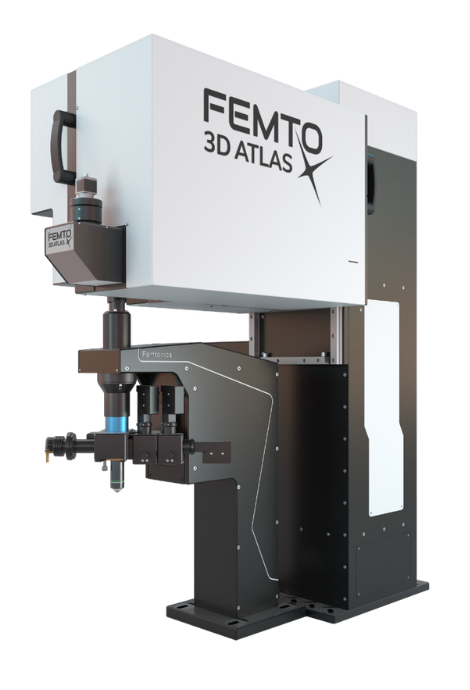
With the truly 3D, blazing-fast Real-Time Motion Correction solution, Femtonics FocusPinner is available for the acousto-optic FEMTO3D Atlas imaging platform. Explore the unprecedented possibilities in ultra-fast 3D two-photon imaging alongside our growing community of users!
Our solution takes advantage of acousto-optics technology and elevates it to a whole new level. The flexible scanning methods create opportunities to perform correction calculations with a predefined, yet tunable frequency. The incorporation of these measurements has only a negligible impact on our overall measurements, ensuring that our products deliver top-of-the-line speeds even with online motion correction. The algorithm is based on correction calculations derived from selecting stable objects or multiple objects within the field of view (FOV), which serve as reference points during scanning. These reference points can be either injected fluorescent beads or live cells. The correction for the entire FOV is calculated based on the movements exhibited by these areas.
Typical use cases
Neuronal network imaging
Network imaging requires small regions of interests to allow for a reasonable scanning speed. Cells exiting these ROIs generate distortions in the fluorescence data, which can completele change the outcome of an experiment. With motion correction these artifacts can be resolved immediately, to accomodate for a large number of smaller ROIs.
Measurements of long dendritic segments
Corrected motion on structures with a highly irregular shapes such as dendrites can reframe the whole outcome of an experiment. The shape of the activity curve changes substantially by motion artifacts on dendrite, distorting its activity curves, without the motion correction algorithm added to the measurement.
Voltage imaging
As voltage imaging takes the forefront in neuroscientific research, adapting to its high-speed requirements becomes an inevitable step to take. The optimal solution is to measure only the smallest regions of interest (ROIs) possible in order to accelerate the scanning process. However, with small ROIs comes the challenge of motion, which becomes increasingly influential in the measurements as the targets frequently move out of the scanned areas. Furthermore, due to the fast nature of voltage signals, even small errors caused by motion can result in significant aberrations in the data, particularly considering the inherent susceptibility of these signals to aliasing. Therefore, online motion correction is not only useful but also an indispensable tool in voltage imaging.
Do you think our cutting-edge devices could boost your research projects?
|
FEMTO3D Atlas: Breakthrough innovation in two-photon microscopy |
