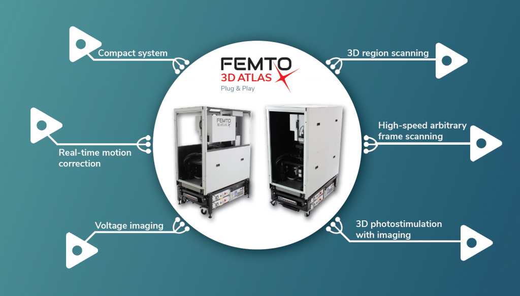
What is FEMTO3D ATLAS PLUG & PLAY?
The FEMTO3D ATLAS PLUG & PLAY microscope is a turnkey multiphoton solution: following a smooth delivery to the laboratory, it is ready to operate within an hour. The system can be easily relocated or moved between laboratories, adapting to your ever-changing needs. While compact in size, the microscope is equipped with the latest 3D acousto optic (AO) technology for ultra-fast in vivo 3D imaging and 3D photostimulation.
The FEMTO3D ATLAS PLUG & PLAY combines high tech science and engineering in 3D measurements. It performs and goes beyond all that galvo and resonant scanner based imaging can do and extends into three dimensions, providing the all-in-one solution in two-photon microscopy.
Ultra-fast, simultaneous 3D imaging and 3D photostimulation in a large volume with real-time motion correction: capture dendrites and whole networks at the same time! Scan silently, without mechanical restrictions!
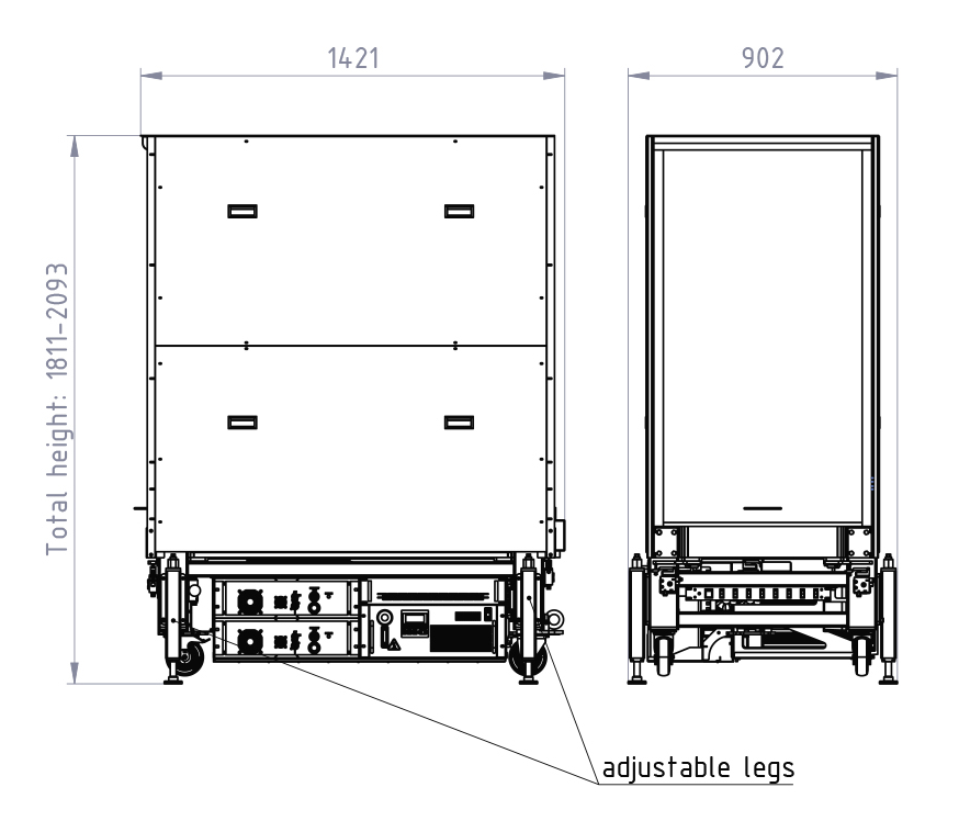
ROLLS OUT OF THE BOX
Compact and arrives ready to operate
FUNCTIONAL REAL-TIME 3D IMAGING
Calcium imaging, voltage imaging
DEEP PENETRATION
Low phototoxicity, high optical quality
UNIQUE FLEXIBLE IMAGING METHODS
Supporting neurobiological applications
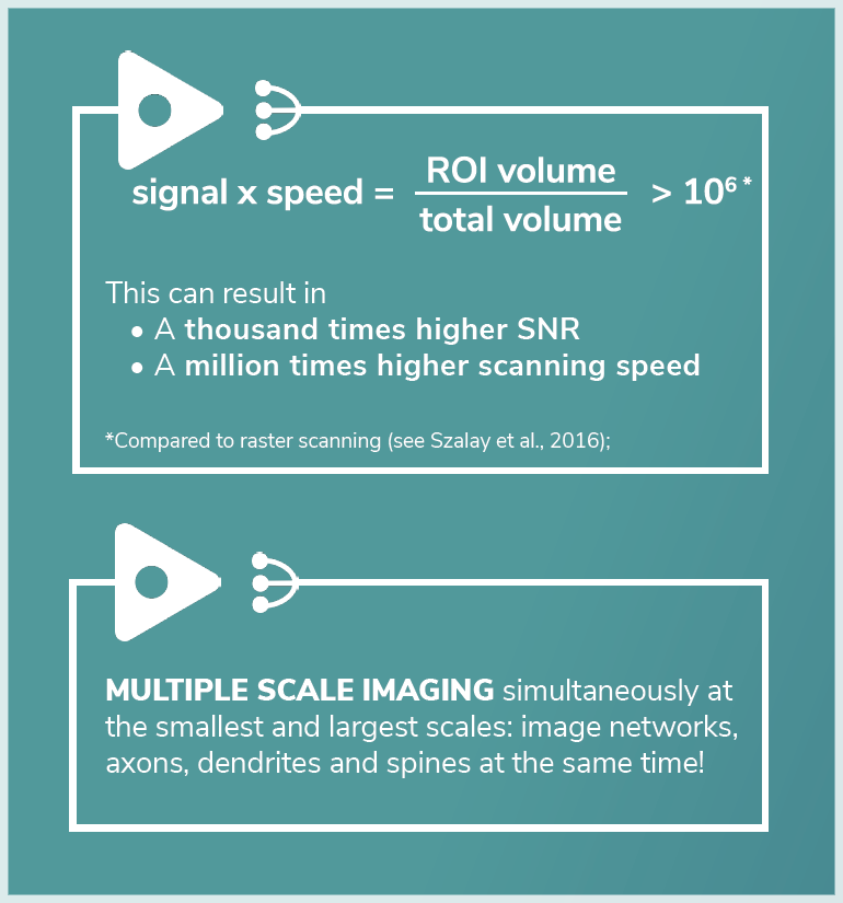
NETWORK IMAGING
Of over 2000 soma distributed in 3D
DENDRITIC IMAGING
And spine mapping without interruption
DURING BEHAVIOR
Real-time 3D motion correction
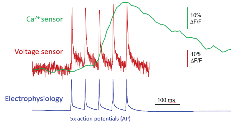
VOLTAGE IMAGING
The rapidly evolving voltage sensor technology determines the next decade of bio-imaging: they have faster kinetics than calcium sensors and can provide superior temporal resolution, capable of detecting spikes at frequencies greater than 100 Hz. Combined with the high sampling rate of the Atlas Plug & Play (up to 100kHz), they are suited for detecting ultrafast transients, such as action potentials (APs). The AO technology also surmounts mechanical distortion and jumping delay between ROIs, spending recording time only at structures of interest. The Atlas Plug & Play powerd by the AO technology, with real-time motion correction, is currently the only imaging device which can keep up with the speed of
firing neurons.
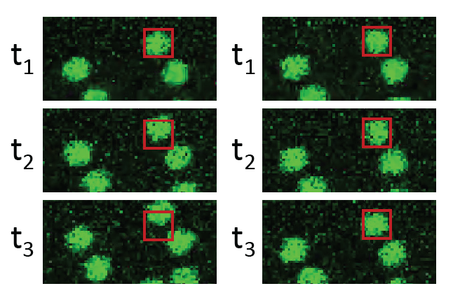
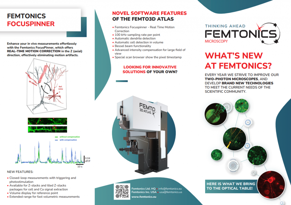
REAL-TIME 3D MOTION CORRECTION
Drawing reliable conclusions from data collected from behaving animals can be challenging, as artifacts arising from tissue movements can be difficult to discern from real activity signals. The Femtonics FocusPinner real-time 3D motion correction feature has been developed to eliminate motion artifacts during data acquisition not just along the axes of the imaged plane but also along the optical axis. By combining it with the scanning modes of the Atlas Plug & Play, neuronal activity data can be aquired without motion artifacts while the animal is performing tasks in virtual reality. FocusPinner will allow you to profit from unprecedented signal-to-noise ratio and save time by avoiding post-hoc processing.
ABOUT THE FEMTO3D Atlas Plug & Play
• In vivo functional imaging down to below 850 µm depth
• 500 µm × 500 µm × 650 µm scanning volume in vivo (with a 20x, NA=1.0 obj.)
(In case of excellent labelling it can be exteded to 800 µm x 800 µm x 1050 µm while keeping the good central resolution)
• Wavelength between 920 – 1040 nm
• Integrated automatic beam stabilization
• Integrated dispersion compensation unit for most effective excitation
• Diffraction limited, submicrometer resolution in the center (<450 nm)
• Scanning speed up to 100 kHz to any points in 3D
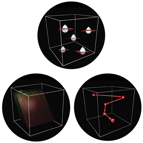
• 3D SCANNING MODES: random-access point, trajectory, tilted frame, volume, ribbon, snake, chessboard, multi-cube scanning
• Quasi-simultaneous 3D imaging and 3D photostimulation
• Real-time 3D Motion Correction to eliminate motion artifacts arising from tissue movements
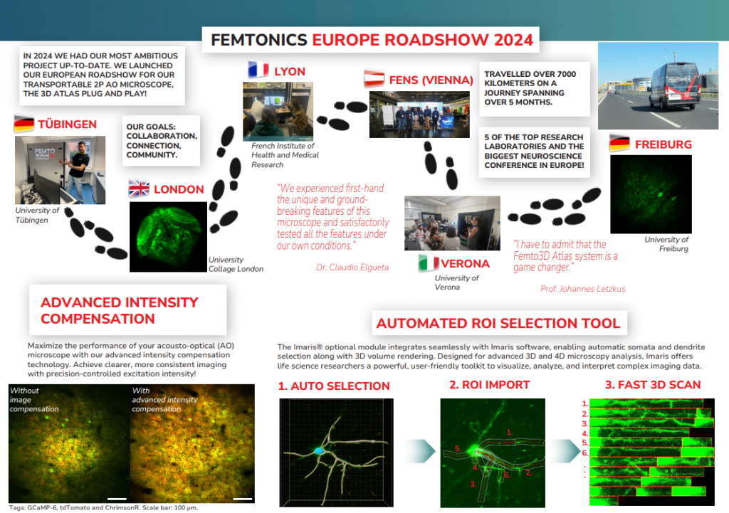
Plug & Play European Tour 2024
What a journey it’s been! We had an amazing time traveling across Europe with our FEMTO3D Atlas Plug&Play microscope, and it was a true pleasure to connect with so many of you along the way. Experiencing your enthusiasm and seeing the PnP system in action made this tour unforgettable.
A big thank you to everyone who joined us, shared insights, and helped make this event a success. Your passion fuels our drive to innovate.
But we’re not done yet! We’re already planning for even bigger things next year. Until then, stay curious and stay tuned—because we’re looking forward to seeing you again in 2025!
Would you like to learn more?
Get in touch with our experts and learn more about the special applications of the FEMTO3D Atlas Plug & Play.