Genetically encoded voltage indicators (ASAP, JEDI2P, etc.) have recently attained a level of quality that renders them valuable tools for studying membrane potentials throughout cells. However, a significant challenge has persisted: the lack of sufficiently fast equipment to capture their dynamics. Until now, there has been a shortage of systems capable of imaging at speeds exceeding kHz, approaching the electrophysiological standard of 10 kHz. The Femtonics 3D AO Atlas addresses this gap by enabling high-speed, three-dimensional voltage imaging. Equipped with the real-time motion correction module, the FocusPinner, it achieves rapid imaging without motion artifacts and ensures high signal-to-noise ratio (SNR) for precise voltage imaging.
The rapidly evolving voltage sensor technology is going to determine the next decade of neuroscience. Voltage signals have faster kinetics and thus provide and require superior temporal resolution. The sensors report individual action potentials even within fast bursts. Conventional imaging techniques are not fast enough to capture these transients.
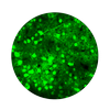
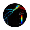
Figure 1: Visualizing the speed of the signal and the proof of necessity for Acousto Optical scanning technology. A) Green: Ca2+ from a PV interneuron sampled at 30 Hz Red: JEDI2p sampled at 8.4 kHz. Blue: Patch clamp electrophysiological recording of 5 evoked Action Potentials sampled at 20 kHz. B) Same measurement down–sampled to 80, 40 and 21 Hz.
One of today’s most interesting and challenging task is to measure voltage signals from neurons while the model animal is active. Even recently published in vivo voltage imaging studies have several limiting factors. Femtonics has overcome these limitations and allowed researchers to have higher success rate, higher SNR, higher number of recording locations in 3D, better focus on the dendrites and alternative ways to investigate and interact with neuronal population. All measurement techniques can be easily applied in head-fixed behaving animals too, due to the 3D Real-Time Motion Correction, which enables the high SNR in vivo as well.
Attila Losonczy, MD, PhD – Professor of Neuroscience; Principal Investigator at Columbia’s Zuckerman Institute
Figure 2: A) 3D model showing the relative locations of the measured neurons. B) Quality JEDI2P labelling in the visual cortex L2/3. C) Example traces showing the SNR with 1 kHz sampling from a behaving animal. Bottom: Chessboard scanning of 5 individual neurons from different Z planes. D) Average of individual APs. (n=17)
Figure 3: A) Chessboard ROIs in 600 x 600 x 600 um volume B) ASAP labelling in the in M1 region, red squares mark the recorded areas with low resolution. C) Voltage traces from 13 neurons in head-fixed conditions recorded with 400 Hz speed.
Figure 4: A) 3D model showing a chessboard and ribbon scans from the same neuron in visual cortex B) Recorded ROIs from different planes. C) Voltage traces the recorded areas D) Soma and the two different dendritic segment aligned.
Figure 5: A) Experimental setup: mice were active on a treadmill while undergoing two-photon voltage imaging with Femto3DATLAS. Auditory stimulus served as activation signal in the V1 cortex. B) 3D model of the network which was recorded with line scans. C) JEDI2P labelled neurons in the field of view. Red lines mark the location of the line scans with 2.2kHz D) Averaged signal responses for the auditory stimulus from 17 neurons. Green: jRGECO calcium signals, Red: JEDI2P voltage signals excited on 1040nm. The black line shows the timing of the auditory stimulus.
Figure 6: A) Schematics of Drosophila 2p imaging. B) ASAP labelled Drosophila neurons. Red lines over the somata are representing the line scan locations C) ASAP signal from the labelled neuron recorded with 6675.57 Hz.
We offer optical electrophysiology solutions for problems that are impossible or close to impossible to solve with classical electrophysiology (i.e. multiple site dendritic patch, repeated bouton patch etc.) working today in the patch clamping world of electrophysiology and photoactivation, as multiple measurements sites can be recorded with up to 100 kHz by applying the DRIFT scanning in 3D. (Szalay et al. 2016)
Dr. Christopher D. Makinson – Assistant Professor of Neurological Sciences (in Neurology, and the Institute of Genomic Medicine Columbia University).
Figure 7: Validation of concept comparison of acousto-voltage imaging and electrophysiology measurements. A) Whole cell patch–clamp camera image and the maximum intensity projection of the patched neuron under two–photon microscopy B) Rise time comparison of the JEDI2P and the electrophysiological signal C) Half-width on the top and signal amplitude measurement comparisons on the bottom.
Figure 8: Combined two-photon and electrophysiology of HEK 293 cells. A) Camera image and fluorescent labelling of the field-of-interest. JEDI2P in the green channel. The measurements were done along the line marked with red. B) Fluorescent intensity change during ramp test on a HEK cell. The intensity decreased under the detection threshold C) Top is raw data, bottom is filtered. Somatically evoked ramp test. Blue: electrophysiology, red is voltage signals measured with 8.32 kHz.
Figure 9: A) 3D model of a single pyramidal neuron measured in vitro with 3D line scan. The red mark is showing the recorded region with a 3D line scan. B) Maximum intensity Z projection of a cortical pyramidal cell. White dots are indicating the measurement locations. C) Somatically evoked 5 backpropagating action potentials. Left: Fluorescent intensity map of JEDI2P from pyramidal cell’s apical dendrite. Right: Temporal integration of the intensity map at the given ROI. There were 24 ROIs from the measured dendritic area with 2.24 kHz. Bottom: Arrows indicating the somatically evoked events, blue: electrophysiology, red: integrated curves for the whole dendrite based on the JEDI2P signal.
Figure 10: A) 3D reconstruction of a dendrite with the uncaging locations marked with yellow. The imaging as 3D line scan marked with blue B) Maximum Z-projection of a pyramidal neuron (ALEXA-594) and in the inset the scanning line (blue), uncaging locations (yellow, 15 locations, 0.3 ms ROI time). C) Examples of uncaging (DNI-Glu) evoked EPSP (top, JEDI-2P signal on dendrite, bottom somatically recorded electrophysiological signal).
Coming soon:
Reference labs are located at: Stanford University , Baylor College of Medicine, Columbia University, Basel University, NIH and BrainVisionCenter.
We are eager to demonstrate you our capabilities online in vitro or in vivo. Book an appointment to see how easy to achieve such results.
More developments are in the works that focus on voltage imaging such as disc scanning, 3-level image stabilization, immediate multi-channel analysis, etc.
Our partners have an extremely high grant success rate with voltage imaging. We are ready to help you and work with you on your grant applications: CONNECT to you sales representative for grant application assistance!
All sensations and behaviors are encoded in dynamic activity patterns of neural networks. In other words, complex structures of many individual neurons responding to environmental features, visual or auditory stimuli,, reward or punishment, etc. These neural systems extend over 3D space and, in most cases, cross many cortical layers of the brain. With two-photon microscopy enabling neuroscientists to reach the deep regions of the brain (down to 850 µm), it is now possible to study neural networks at a high spatiotemporal resolution.
The FEMTO3D Atlas microscope with its unique technical solutions enables the precise spatial and real-time complexity of neuronal coding to be resolved by scanning large number of cells distributed in a 3D volume.
Several thousand cells can be measured near-simultaneously (up to 100 kHz per point) by 3D random-access point scanning by restricting the imaging to sub-regions of the 3D volume, which results ultra-high scanning speed and signal-to-noise ratio.The figure (A-D) shows the changes in Ca2+ concentration of 2000 cells from the visual cortex of a GCaMP-expressing mouse as a function of time. See more:
Wertz et al., Science, 2015; Katona et al., Nat Meth, 2012.
Chessboard scanning is a planar extension of random-access point scanning, where random-access points are extended to small squares by drifting the laser beam. These squares can be located anywhere in the imaging volume and include the somata with the surrounding area. Up to 300 somata can be measured simultaneously. The chessboard arrangement of the squares helps to visualize somata, analyze their activity, and correct for motion artifacts, and completely eliminating them with the help of the Femto FocusPinner. The video shows in vivo neuronal activity, Ca2+ transients from 100 neuronal somata from the mouse V1 region, labeled with GCaMP6. See also Szalay et al., Neuron, 2016.
Sparsely distributed brain cells (such as interneurons) can be followed in real-time in a 3D volume of using chessboard scanning. In the study depicted in the figure below, the calcium activity from nearly 250 interneurons was measured simultaneously, across every CA1 hippocampal layer, in an 800 × 800 × 500 µm volume with the chessboard scanning mode, while the mouse was performing behavioral tasks on a treadmill. Using these recordings, the biochemical composition of the imaged cells was also determined from data acquired from post hoc immunohistochemistry. Such a novelty – identifying distinct interneuron types and their activity dynamics simultaneously in a large neural network – could not have been achieved with other traditional recording techniques. See more: Geiller et al., Neuron, 2020. Watch the interview with one of the authors.
Multi-cube scanning is a spatially extended mode of chessboard scanning, where a Z dimension is added to the chessboard squares to cover the whole extent of the somata, therefore preserving all fluorescent information from the somata even during large amplitude movements Learn more: Bharioke et al. Neuron (2022)
Our solution takes advantage of acousto-optics technology and elevates it to a whole new level. The flexible drift scanning method creates the opportunity to perform correction calculations with a predefined and adjustable frequency. The algorithm is based on correction calculations derived from selecting stable objects within the field of view (FOV), which serve as reference points during scanning. With our method providing an effortless means to scan in any direction in 3D, reference measurements are equally efficient in the Z direction as they are in the XY direction, which is a unique feature of the Femtonics FocusPinner.
Cells assembled into a neural network can be acquired with a frame rate of 40 Hz in a 500×500 µm field-of-view (FOV) by the High-speed arbitrary frame scanning mode of the Atlas. Beyond the high speed, another important and unique advantage of this mode is that the selected plane can be rotated into any in space to match your layer of interest.
The video below demonstrates the rotation of the selected plane in High-speed arbitrary frame scan mode. The left panel shows the relative position of the objective’s focal plane with blue and the actual two-photon focal plane with pink. The movement of the pink plane exemplifies the possibilities Atlas offers you to orient the plane in a three dimensional tissue. The right panel shows the neurons of a network in the actual scanned plane during rotation in live mode.
FEMTOSmart offers numerous imaging opportunities and with its vast modularity researchers can study neuronal networks from varied perspectives and save time in the experimental work.
The FEMTOSmart Galvo, with its flexible scanning patterns such as 2D random-access point, 2D multiple-line, and folded-frame scanning, supports the manual selection of individual cells in a neural network in a 2D plane. By skipping the measurement of the entire field, it is possible to reach a fast scanning speed and maintain a high signal-to-noise ratio. Using the TravellingSalesman additional software module, it is possible to determine the shortest pathway visiting defined points arbitrarily dispersed in the field of view. The short round-trip time results in a higher measurement repetition speed, up to 100 Hz for about 30 cells.
Fast-frame scanning by FEMTOSmart Resonant combined with fast Z-focusing performed by a Piezo objective positioner kit is a well-known approach for studying any three-dimensional neural network. In this case, the entire field of view is imaged continuously by the fast scanner, while the objective positioner moves between planes. More than one (cortical) layer might be recorded simultaneously this way, or the frames may be assembled into volumes resulting in a four-dimensional set of data.
Cortex-wide response mode of VIP-expressing inhibitory neurons by reward and punishment Zoltán Szadai, Hyun-Jae Pi, Quentin Chevy, Katalin Ócsai, Dinu F Albeanu, Balázs Chiovini, Gergely Szalay, Gergely Katona, Adam Kepecs (2022)
Large-Scale 3D Two-Photon Imaging of Molecularly Identified CA1 Interneuron Dynamics in Behaving Mice Tristan Geiller, Bert Vancura, Satoshi Terada, Eirini Troullinou, Spyridon Chavlis, Grigorios Tsagkatakis, Panagiotis Tsakalides, Katalin Ócsai, Panayiota Poirazi, Balázs J. Rózsa, Attila Losonczy / Neuron (2020)
Cell Types of the Human Retina and Its Organoids at Single-Cell Resolution Cell Types of the Human Retina and Its Organoids at Single-Cell Resolution Cameron S. Cowan, Magdalena Renner, Martina De Gennaro, Brigitte Gross-Scherf, David Goldblum, Yanyan Hou, Martin Munz, Tiago M. Rodrigues, Jacek Krol, Tamas Szikra, Rachel Cuttat, Annick Waldt, Panagiotis Papasaikas, Roland Diggelmann, Claudia P. Patino-Alvarez, Patricia Galliker, Stefan E. Spirig, Dinko Pavlinic, Nadine Gerber-Hollbach, Sven Schuierer, Aldin Srdanovic, Marton Balogh, Riccardo Panero, Akos Kusnyerik, Arnold Szabo, Michael B. Stadler, Selim Orgül, Simone Picelli, Pascal W. Hasler, Andreas Hierlemann, Hendrik P. N. Scholl, Guglielmo Roma, Florian Nigsch, Botond Roska (2020)
Fast 3D Imaging of Spine, Dendritic, and Neuronal Assemblies in Behaving Animals. Gergely Szalay, Linda Judak, Gergely Katona, Katalin Ocsai, Gabor Juhasz, Mate Veress, Zoltan Szadai, Andras Feher, Tamas Tompa, Balazs Chiovini, Pal Maak, Balazs Rozsa, Neuron (2016)
Single-cell-initiated monosynaptic tracing reveals layer-specific cortical network modules Wertz A, Trenholm S, Yonehara K, Hillier D, Raics Z, Leinweber M, Szalay G, Ghanem A, Keller G, Rozsa B, Conzelmann KK, Roska B, Science (2015)
Fast two-photon in vivo imaging with three-dimensional random-access scanning in large tissue volumes Gergely Katona, Gergely Szalay, Pál Maak, Attila Kaszas, Mate Veress, Daniel Hillier, Balazs Chiovini, E Sylvester Vizi, Botond Roska & Balazs Rozsa, Nature Methods (2012)
Dendrites and dendritic spines are made of fine, thin and elongated segments therefore they are difficult to study. However, using two-photon technology, we are able to collect signals from deeper regions of the brain, while avoiding phototoxicity. Additionally, spatially confined, regional scanning of axons, dendrites and spines allows detecting even subthreshold signals with high signal-to-noise ratio (SNR). We have several configurations aiding to visualize dendritic arborization, and perform functional measurements in vivo and in vitro. Now, with the help of our new real time motion-correction called Femtonics FocusPinner motion artifacts even in vivo in dendritic measurements will no longer be a problem!
With the FEMTO3D Atlas point scanning speeds up to a 100kHz can be achieved, which helps determining activity at virtually any sites on dendrites. 3D random-access point scanning and its advanced versions support all kinds of functional dendritic imaging from single spine investigations to full arborization measurements, even combined with photostimulation solutions.
The video shows a 3D recording of Ca2+ signals at multiple dendritic sites by 3D random-access point scanning. See more in Chiovini et al., Neuron, 2014.
3D random-access point scanning extended by drifting the focal point along short 3D trajectories allows imaging without interruption on multiple, long dendritic branches. The sampling is continuous during the drift, so this scanning mode gives a more detailed spatial resolution without changing the overall scanning time. As a result, the function of thin dendritic segments, or even spines can be determinded, and single action potentials can be observed. See more in , Chiovini et al., Neuron, 2014.
An extension of the 3D multiple-line scanning is 3D ribbon scanning, which makes it possible to image ribbon shaped surfaces containing dendrites and the neighboring areas. Figures show 3D ribbons encompassing seven dendritic segments with their spines of a GCaMP6-labeled layer II/III pyramidal neuron measured within the brain of a living mouse.
The seven ribbons were projected into a 2D image ordering dendrites above each other for better visualization, and activity was recorded from 40 selected spines and visualized in the form of classical Ca2+ transients.
Our solution takes advantage of acousto-optics technology and elevates it to a whole new level. The flexible drift scanning method creates the opportunity to perform correction calculations with a predefined and adjustable frequency. The algorithm is based on correction calculations derived from selecting stable objects within the field of view (FOV), which serve as reference points during scanning. With our method providing an effortless means to scan in any direction in 3D, reference measurements are equally efficient in the Z direction as they are in the XY direction, which is a unique feature of the Femtonics FocusPinner.
High-speed arbitrary frame scanning gives you the freedom to tilt and rotate the scanning plane in any direction. With this feature, in vivo imaging of long dendrites can be performed in a single plane. To examine multiple neurons’ apical dendrites simultaneously, the tilted frame can be extended into a volume element.
The video shows the apical dendrites of the neurons getting aligned with the scanning plane, which means the spatio-temporal activity patterns of these long segments passing multiple cortical layers can be recorded without requiring lengthy and painstaking optical sectioning.
FemtoSmart offers numerous imaging opportunities and thanks to its modularity researchers can study dendrites with spines from wide variations of perspectives.
The adaptable X and Y mirrors of the galvanometric scanner, coupled with the special electronic boards from Femtonics, can follow the tortuous protrusions of the dendritic arbor precisely using the 2D random-access point, multiple-line and folded frame scanning methods. By limiting the scanning to the interesting dendritic segments carrying spines and omitting the space between them, both the scanning speed (up to 2 kHz) and the SNR can be increased using 2D random-access point and multiple-line scanning modes. The advantage is multiple-line scanning is the imaging without interruption at multiple dendritic branches, it is the cost-effective, alternative solution compared to the acousto-optic scanner-based line scanning methods.
Using folded frame scanning, an area along a pre-selected line can be imaged. The selected regions can take many shapes, from areas around straight lines to complex bent curves. This advanced scanning method is useful for following events along curved dendrites with spines, and can also be advantageous for dendritic measurements in behaving animal models where motion artefacts are a common problem. The images are corrected for motion offline by the control software, as long as the dendrite remains in the scanned area.
As dendritic segments are abundant, complex, 3 dimensional features of the brain, selecting ROIs around them is a maticulous job, reliant on human perception. With the help of this optional module, you will be able select all dendritic segments in your field of view, just with some push of buttons. This will save you time and energy, and the end result will be more precisely aligned, down to the pixel level!
Pyramidal neurons form active, transient, multilayered circuits perturbed by autism-associated mutations at the inception of neocortex Munz M, Bharioke A, Kosche G, Moreno-Juan V, Brignall A, Rodrigues TM, Graff-Meyer A, Ulmer T, Haeuselmann S, Pavlinic D, Ledergerber N, Gross-Scherf B, Rózsa B, Krol J, Picelli S, Cowan CS, Roska B
Fast 3D Imaging of Spine, Dendritic, and Neuronal Assemblies in Behaving Animals. Gergely Szalay, Linda Judak, Gergely Katona, Katalin Ocsai, Gabor Juhasz, Mate Veress, Zoltan Szadai, Andras Feher, Tamas Tompa, Balazs Chiovini, Pal Maak, Balazs Rozsa, Neuron (2016)
Dendritic spikes induce ripples in parvalbumin interneurons during hippocampal sharp waves. B Chiovini, G F Turi, G Katona, A Kaszas, D Palfi, P Maak, G Szalay, M F Szabo, Z Szadai, Sz Kali and B Rozsa, Neuron (2014)
Fast two-photon in vivo imaging with three-dimensional random-access scanning in large tissue volumes. Gergely Katona, Gergely Szalay, Pál Maak, Attila Kaszas, Mate Veress, Daniel Hillier, Balazs Chiovini, E Sylvester Vizi, Botond Roska & Balazs Rozsa, Nature Methods (2012)
Matching Cell Type to Function in Cortical Circuits. Luc Estebanez, Jens Kremkow, James F.A. Poulet, Neuron (2015)
In Vivo Monosynaptic Excitatory Transmission between Layer 2 Cortical Pyramidal Neurons. Jean-Sebastien Jouhanneau, Jens Kremkow, Anja L. Dorrn, James F.A. Poulet, Cell Reports (2015)
Roller Coaster Scanning reveals spontaneous triggering of dendritic spikes in CA1 interneurons. G. Katona, A. Kaszas, G. F. Turi, N. Hajos, G. Tamas, E.S. Vizi, B. Rozsa, PNAS (2011)
The Central Nervous System organizes behavior, therefore to study CNS functions works best in a behavioral context. In this context it is important to manipulate experimental variables in a precisely controlled way. Those variables may be stimulus information, task instructions, reward or aversive outcome, all affecting the related pattern of brain activation. Monitoring and manipulating behavior together with imaging neural network activity in the awake behaving animal is essential to establish the relation between the cellular/network and behavioral level.
Imaging cellular activity in behaving animals is challenging, since all kinds of movement may generate imaging artifacts additional to that caused by physiological processes like heartbeat and breathing. The presence of physical activity can particularly complicate data collection within the specific timeframe of interest; As the activity of interest is closely related to the movement as behavioral marker, it is very important to be able to keep the recording clean of movement artifacts in a behavioral experiment.
With our truly 3D, blazing-fast motion correction solution, Femtonics FocusPinner, FEMTO3D Atlas imaging platform can follow the displacement of the ROIs during the experiments eliminating artifacts caused by heartbeat, breathing or physical activity allowing calcium and voltage imaging from dendrites and somata with high resolution.
Residual displacements can be corrected by post-hoc movement correction techniques.
Adjacent drifts may cover surface-like recording regions, surfaces can be extended to form volumes. This way areas or volumes around the objects of interest can be recorded thereby providing basis for a post-recording motion correction allowing to preserve physiological data despite tissue movements. (Szalay et. al, Neuron (2016). Post-hoc movement correction algorithms built in the software ensure subresolution alignment of ROIs even at large artifacts in 3D.
The surface scanning methods are optimized for speed, while the methods based on volume imaging are optimized for large amplitude movements. Each scanning mode is useful for different neurobiological aims: ribbon scanning, snake scanning, 3D multiple line scanning areis optimal for various dendritic measurements, while chessboard scanning and multi-cube scanning are best used for somatic recordings.
Learn more about the 3D Anti-motion technology and fast 3D AO imaging in behaving animals from Szalay et. al, Neuron (2016)
The top panel shows the schematic diagram of the experiment. Two-photon imaging was performed using the 3D Multiple-line scanning mode of the FEMTO3D Atlas. At specific locations, the animal received a reward (reward zone) or an unpleasant stimulus (aversive zone). The bottom panel shows the selected neurons and location of imaging and their calcium activity aligned with the collected motion data. Aversive and reward zones are marked with red and green, respectively.
Left panel: schematic diagram of the experiment. VIP interneurons were imaged by the Chessboard scanning mode of the FEMTO3D Atlas, while the mouse was performing an auditory discrimination task. In this “classical” behavioral experimental protocol, water-restricted mice were trained to associate water reward with a „go” tone, and punishment with a „no-go” tone. Altogether 811 VIP cells were imaged from 18 brain regions. On the right panel, each row of the raster plot represents the average neuronal response for Hit and False alarms, and the graph shows the Ca2+ responses of one VIP interneuron averaged for Hit (thick green), False alarm (thick red), Miss (thin green), and Correct rejection (CR, thin red).
Learn more: Szadai et. al 2022
In these behavioral tests, water restricted mice were trained to run on a fabric belt enriched with tactile cues, and to operantly lick and receive water rewards depending on the type of the experiment. The velocity of the animal was recorded, and hippocampal interneurons were imaged simultaneously by the Chessboard scanning mode of the FEMTO3D Atlas.
The figure shows that more than 200 interneurons were imaged simultaneously during the random foraging behavioral task, and the locomotion state of the animal (velocity, locomotion, immobility) was associated with the Ca2+ activity of the subsequently characterized interneuron types.
Learn more: Geiller et al. Neuron (2020)
The FEMTOSmart product line was primarily developed to perform in vivo imaging. This system’s special feature is the elevated body that can move in X, Y, and Z dimensions: it offers plenty of space under the objective for free and easy positioning of any kind of sample. This feature supports functional behavioral studies of model organisms with different weights and sizes, from flies to even non-human primates, while they are moving in a virtual-reality environment. The microscopes can be assembled using galvanometric scanner supporting flexible ROI scanning and resonant scanner supporting fast frame and volume scanning possibilities. Many optional modules are available to the microscopes enabling the scientists to perform optogenetics and other studies in larger animals even during behavior.
Dal Maschio et al. described a method for high-resolution interrogation of circuit function, which combines simultaneous two-photon functional imaging, 3D photostimulation, and quantitative behavioral tracking in zebrafish larvae. Tail movements were initiated by photostimulation, while the induced behavior and neuronal network activity were recorded. The schematic diagram of the experiment is shown on the left panel of the figure. The right panel shows the calcium activity of 486 imaged neurons and the kinematics of the tail in response to stimulation. Red lines indicate photostimulation onset and offset.
Learn more: dal Maschio et al. Neuron (2017)
Böhme et al. used two-photon imaging to record calcium responses during a short-term memory test in Drosophila. In this classical aversive olfactory conditioning experiment, flies were trained by pairing an odor with an electric shock. During the simultaneous two-photon imaging and behavioral experiment, calcium activity was evoked in the neurons by odors. See the left panel of the figure for the schematic diagram. On the right panel, averaged odor responses are shown in control and mutant Drosophila. Five responses per odor were averaged per animal.
Learn more: Böhme et al. Nat Commun (2019)
Head holder stands and head plates fix the rodent’s head in various positions, adapted to the conditions and enabling precise measurements in the brain, the eye and other parts of the head. Two types of head holders are available with different properties for different purposes, ie. recording from anesthetised or behaving animals etc.
Bpod is an open-source behavior-recording system and real-time environment controller for rodent experiments. Using high-level programming environments (MATLAB/Python), it provides a low-latency closed-loop link between behavioral events, stimulus delivery, and stimulation. Using its built-in liquid reward delivery system, it can be used to power go/no-go discrimination, two alternative forced choice, and CS/US behavioral paradigms. Bpod data acquisition is fully synchronized with the microscope, and the behavioral events can be precisely aligned to the recording of Ca2+ transients.
Our microscopes have a wide range of high sampling rate communication capabilities (analog and digital input/outputs) which support a high level of synchronization with various external devices. This includes triggering either ways or generating stimulation or stimulation patterns. Visual, auditory, tactile, whisker or odor stimuli can be generated this way. The MES command line access and FemtoAPI allow custom scripting of the measurement control including third party devices to form a complex environment for behavior experiments.
Cortex-wide response mode of VIP-expressing inhibitory neurons by reward and punishment Zoltán Szadai, Hyun-Jae Pi, Quentin Chevy, Katalin Ócsai, Dinu F Albeanu, Balázs Chiovini, Gergely Szalay, Gergely Katona, Adam Kepecs eLife (2022)
Sharp-wave ripple doublets induce complex dendritic spikes in parvalbumin interneurons in vivo Linda Judák, Balázs Chiovini, Gábor Juhász, Dénes Pálfi, Zsolt Mezriczky, Zoltán Szadai, Gergely Katona, Benedek Szmola, Katalin Ócsai, Bernadett Martinecz, Anna Mihály, Ádám Dénes, Bálint Kerekes, Áron Szepesi, Gergely Szalay, István Ulbert, Zoltán Mucsi, Botond Roska & Balázs Rózsa, Nature Communications (2022)
Large-Scale 3D Two-Photon Imaging of Molecularly Identified CA1 Interneuron Dynamics in Behaving Mice Tristan Geiller, Bert Vancura, Satoshi Terada, Eirini Troullinou, Spyridon Chavlis, Grigorios Tsagkatakis, Panagiotis Tsakalides, Katalin Ócsai, Panayiota Poirazi, Balázs J. Rózsa, Attila Losonczy, Neuron (2020)
Fast 3D Imaging of Spine, Dendritic, and Neuronal Assemblies in Behaving Animals. Gergely Szalay, Linda Judak, Gergely Katona, Katalin Ocsai, Gabor Juhasz, Mate Veress, Zoltan Szadai, Andras Feher, Tamas Tompa, Balazs Chiovini, Pal Maak, Balazs Rozsa, Neuron (2016)
Accurate spike estimation from noisy calcium signals for ultrafast three-dimensional imaging of large neuronal populations in vivo. Thomas Deneux, Attila Kaszas, Gergely Szalay, Gergely Katona, Tamas Lakner, Amiram Grinvald, Balazs Rozsa & Ivo Vanzetta, Nature Communications (2016)
Microglia protect against brain injury and their selective elimination dysregulates neuronal network activity after stroke. Szalay G, Matinecz B, Lenart B, Kornyei Z, Orsolits B, Judak L, Csaszar E, Fekete R, West BL, Katona G, Rozsa B, Denes A, Nature Communications(2016)
Matching Cell Type to Function in Cortical Circuits. Luc Estebanez, Jens Kremkow, James F.A. Poulet, Neuron (2015)
Three-photon (3P) microscopy allows noninvasive structural and functional imaging by making cells visible in deep tissues with high spatial resolution and better contrast compared to two-photon excitation. The optical design required for 3P excitation establishes higher axial resolution than that of two-photon microscopy. The spectral window enables three-photon excitation of a variety of fluorophores, such as the current generations of protein-based genetically encoded calcium indicators (e.g. GCaMP6) and the repetition rate of the laser source is adequate for imaging Ca2+ transients produced from neural activity.
The video shows an approximately 1000 μm thick 3P z-stack from a mouse cortex. Purple: blood vessels, green: GFP labelled pericytes. Footage courtesy of Dr. Severin Filser.
3P range microscopy
FEMTOSmart Galvo equipped with 3P range microscopy optional module allows excitation by a wavelength range of 1,200-1,700 nm. The longer excitation wavelengths are scattered less in the biological tissues, which allows extending the penetration depth, reduces out-of-focus excitation, and increases the signal-to-noise ratio. The figure, prepared using a FEMTOSmart Galvo with a 3P range microscopy module, shows spontaneous neuronal activity in the GCaMP6f-labeled visual cortex of a mouse: the cells were excited using a 1300 / 1400 nm laser wavelength for 3P excitation.
Third Harmonic Generation is a specific effect of 3P excitation, that results from the conversion of three incoming photons into one emitted photon with tripled energy and thus, the emission of the light of one-third the wavelength. THG occurs at structural interfaces that are formed between aqueous fluids and lipid-rich structures, for instance, biological membranes, and between water and large protein aggregates such as collagen bundles or muscle fibers.
The figure shows a THG image of a section of a mouse kidney: kidney cells were excited at 1500 nm, and the emitted photons were collected at the green channel (~500 nm) of the detector system.
“We have found a massively great partner in Femtonics and acquiring the groundbreaking 3P (three-photon) FEMTO SMART system ensures never-seen experimental results in deep imaging. We are impressed how fast they are reacting at our requests and able to think together with us on scientific solutions. Their technical level and support are professional, and it is always good to have a trustful partner in daily scientific life.”
Dr. Severin Filser
DZNE – German Center for Neurodegenerative Diseases, Germany
Fluorescence-lifetime imaging microscopy (FLIM) delivers both structural (intensity image) and functional (lifetime image) information and minimizes the imaging effect of photon scattering in thick layers of sample. FLIM provides an accurately quantitative assessment of changes of many biophysical parameters in a target molecular microenvironment, without being affected by the intensity of the emission, quenching or uneven fluorophore distribution. The most frequent FLIM applications are the measurement of molecular environment parameters, molecular interaction and distance measurements by Förster resonance energy transfer (FRET), besides studying the metabolic state of cells and tissues via their autofluorescence. Two-photon fluorescence-lifetime imaging microscopy (2P-FLIM) meets the needs of neuroscientific research because of the combined advantages of two-photon excitation and fluorescence-lifetime imaging: superior depth penetration and decreased photo-bleaching, owing to long excitation wavelengths, and inherent optical sectioning.
The FEMTOSmart Galvo upgraded with the FLIM extension is suitable for performing in vivo and in vitro 2P-FLIM studies in a wide range of model organisms. In this complex system, up to two hybrid detectors (combination of GaAsP cathode and an avalanche detector) with two photon counting channels provide a high detection efficiency and a fast timing response, with less noise. As afterpulsing is essentially absent in these detectors, the dynamic range of fluorescence decay recordings is considerably increased. The special scanning modes and patterns of the FEMTOSmart Galvo provide high spatiotemporal resolution during measurements. The setup is perfectly suitable to monitor glutamate release and intracellular calcium concentration, to perform FRET measurements, or to quantify the ratio between reduced and oxidized NADH and FAD molecules, in the research of neurodegenerative diseases or to reveal metabolic changes during tumorigenesis in cancer research.
A FEMTOSmart Galvo with a FLIM extension is suitable to follow single action potential-evoked presynaptic Ca2+ dynamics, with nanomolar sensitivity, in individual small, presynaptic boutons in acute brain slices. Jensen et al., from the laboratory of Prof. Dmitri Rusakov, have advanced a two-photon excitation FLIM technique. They used the flexible Tornado scanning mode of our galvanometric scanner-based microscope, which provides pixel sampling in a spiral, recording the signal’s spatiotemporal dynamics more favourably, and improves the signal-to-noise ratio. With this method, they performed time-resolved imaging of Ca2+ sensitive fluorescence lifetime of Oregon Green BAPTA-1. In parallel, intensity Tornado imaging made it possible to monitor single-quantum, single-synapse glutamate release in situ directly, using the locally expressed extracellular optical glutamate sensor iGluSnFr. This study paves the way for simultaneous registration of presynaptic Ca2+ dynamics and transmitter release in an intact brain at the level of individual synapses.
(Related article: Monitoring single-synapse glutamate release and presynaptic calcium concentration in organised brain tissue, Thomas P. Jensen, Kaiyu Zheng, Olga Tyurikova, James P. Reynolds, Dmitri A. Rusakov, Cell Calcium, 2017)
FLIM imaging reveals that distinct sites in individual boutons release glutamate in a probabilistic fashion
In a subsequent publication from the same laboratory, Rama et al. describe how they used the FEMTOSmart Galvo with the FLIM extension to monitor spike-evoked glutamate release and presynaptic calcium entry in the mossy fiber boutons of granule cells, with the use of the optical glutamate sensor SF-iGluSnFR and the intracellular Ca2+ indicator Cal-590, and the above mentioned Tornado scanning mode. These presynaptic boutons feature multiple release sites and are densely packed with synaptic vesicles to sustain a highly facilitating “detonator” transmission. In this study, multiplexed imaging showed that distinct sites in individual boutons release glutamate in a probabilistic fashion, while also showing use-dependent short-term facilitation. The results help to reveal whether glutamate release sites act
independently or rather synchronously, and to understand the fundamentals of synaptic transmission and hippocampal network function in a wider context.
(Related article: Glutamate Imaging Reveals Multiple Sites of Stochastic Release in the CA3 Giant Mossy Fiber Boutons, Sylvain Rama, Thomas P. Jensen, Dmitri A. Rusakov, Frontiers in Cellular Neuroscience, 2019)
Fluorescence lifetime imaging reveals regulation of presynaptic Ca2+ by glutamate uptake and mGluRs, but not somatic voltage in cortical neurons. Olga Tyurikova, Kaiyu Zheng, Elizabeth Nicholson, Yulia Timofeeva, Alexey Semyanov, Kirill Volynski, Dmitri A. Rusakov, Journal of Neurochemistry (2020)
Local Resting Ca2+ Controls the Scale of Astroglial Ca2+ Signals. Claire M. King, Kirsten Bohmbach, Daniel Minge, Andrea Delekate, Kaiyu Zheng, James Reynolds, Cordula Rakers, Andre Zeug, Gabor C. Petzold, Dmitri A. Rusakov, Christian Henneberger, Cell Reports (2020)
Multiplex imaging relates quantal glutamate release to presynaptic Ca2+ homeostasis at multiple synapses in situ. Thomas P. Jensen, Kaiyu Zheng, Nicholas Cole, Jonathan S. Marvin, Loren L. Looger, Dmitri A. Rusakov, Nature Communications (2019)
Polymer microchamber arrays for geometry-controlled drug release: a functional study in human cells of neuronal phenotype. Olga Kopach, Kayiu Zheng, Olga A. Sindeeva, Meiyu Gai, Gleb B. Sukhorukov, Dmitri A. Rusakov, Biomaterials Science (2019)
Glutamate Imaging Reveals Multiple Sites of Stochastic Release in the CA3 Giant Mossy Fiber Boutons. Sylvain Rama, Thomas P. Jensen, Dmitri A. Rusakov, Front. Cell. Neurosci. (2019)
A genetically encoded fluorescent sensor for in vivo imaging of GABA. Jonathan S. Marvin, Yoshiteru Shimoda, Vincent Magloire, Marco Leite, Takashi Kawashima, Thomas P. Jensen, Ilya Kolb, Erika L. Knott, Ondrej Novak, Kaspar Podgorski, Nancy J. Leidenheimer, Dmitri A. Rusakov, Misha B. Ahrens, Dimitri M. Kullmann & Loren L. Looger, Nature Methods (2019)
Monitoring intracellular nanomolar calcium using fluorescence lifetime imaging. Kaiyu Zheng, Thomas P Jensen & Dmitri A Rusakov, Nature Protocols (2018)
Monitoring single-synapse glutamate release and presynaptic calcium concentration in organised brain tissue. Jensen TP, Zheng K, Tyurikova O, Reynolds JP, Rusakov DA, Cell Calcium (2017)
Time-Resolved Imaging Reveals Heterogeneous Landscapes of Nanomolar Ca2+ in Neurons and Astroglia. Kaiyu Zheng, Lucie Bard, James P. Reynolds, Claire King, Thomas P. Jensen, Alexander V. Gourine, Dmitri A. Rusakov, Neuron (2015)
Principles of Fluorescence Spectroscopy, 3rd ed. Lakowicz, J.R., Springer (2006)
Fluorescence lifetime maesurement and biological imaging. Berezin, M.Y. & Achilefu, S., Chem. Rev. (2010)
Non-invasive imaging of skin physiology and percutaneous penetration using 5D (space, time and anisotropy) fluorescence spectral and lifetime imaging with multiphoton and confocal microscopy. Roberts, M.S., Dancik, Y., Prow, T.W., et al., Eur. J. Pharm. Biopharm. (2011)
Following blood flow deep in the living tissue
Abnormalities in hemodynamics are linked to various disease states, such as stroke, Alzheimer’s, etc., however, to better understand these associations, more precise optical techniques are needed for interrogating blood flow velocity in vivo in 3D. Acousto-optic (AO) multiphoton microscopy is a superior technique to perform blood flow tracking at the cellular level, at a high spatiotemporal resolution deep in the living 3D tissue. The unique scanning modes of the FEMTO3D Atlas allow you to record continuously along intricate 3D patterns, and its extensions make it possible to follow multiple parameters at the same time, like blood flow and cellular activity. These applications can be useful in various fields, such as neuroscience, immunology, dermatology, oncology and more.
This extension of the FEMTO3D Atlas platform enables fast 3D region of interest (ROI) scanning in a large 3D volume, as well as a quasi-simultaneous detection of neuronal activity and blood flow using two different laser sources. The Femtonics software package provides built-in ROI and curve analysis options, meeting all needs.
Here we injected a red dextran intravenously to make blood vessels appear in red. The passing red blood cells (RBCs) show as shadows in the red stream (Fig1.A). This way, the speed of the blood stream can be measured by following an RBC in a time-dependent manner (Fig 1.B).
Fig 1. How to measure blood flow velocity using 3D two-photon imaging. A. An example image and blood stream visualization from one 3D ribbon in a ~200 um depth. B. A montage image showing the stream of individual red blood cells (yellow circles and arrows). The average speed in a certain part of capillaries or vessels can be calculated from the values of the speed of the red blood cells (v1, v2…vn).
Rapid ROI imaging by the state-of the-art FEMTO3D Atlas AO platform enables 3D ribbon scanning, following vessels through multiple cortical layers in a large brain volume. The total length of the ribbons can reach up to ~5 mm, while maintaining a resolution and speed sufficient to measure the velocity of blood flow (Fig 2.)
Fig 2. Maintaining a proper scanning speed for 3D blood stream measurement, the FEMTO3D Atlas is able to scan an up to 5 mm long ribbon network. A. 3D reconstruction of the ribbon pattern laid down to measure blood flow in mouse cortex. B. Blood stream visualization of a 4.78 mm long 3D ribbon-network; scanning speed: 7 Hz.
Research of cardiac diseases may be supported by a combined method of simultaneous Ca2+ activity and blood flow imaging. This provides a novel opportunity of acquiring local information about neuronal and cardiovascular status. Using the FEMTO3D Atlas Dichro with two laser-sources of different wavelength, blood cell movement and neuronal activity can be measured at the same time. Fast switching between the two laser sources makes it possible to image quasi-simultaneously on two different emission wavelengths. This method can be useful for studying the detrimental consequences of stroke on vascular condition, neuronal damage, and even regeneration processes (Fig 3.).
Fig 3. Simultaneous imaging of neuronal activity and blood flow. Visual cortex neuron appears in green; red indicates a vessel (as a vertical band).
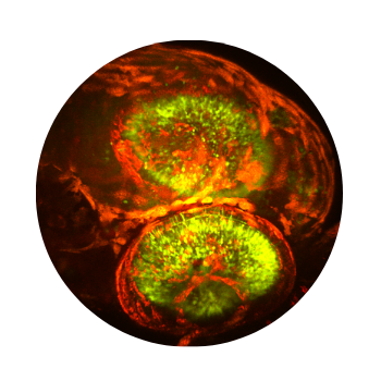
Fig 4. In vivo imaging of blood flow in zebrafish eye. RED: tdTomato labelled blood cells. GREEN: GCaMP6 labelled neurons
The essence of optogenetics is introducing light-activated recombinant ion channels such as channelrhodopsin (ChR2) or halorhodopsin (NpHR) into excitable cells. Light activation of these molecules leads to an influx of ions which induces turning neurons on or off selectively. Halorhodopsin and channelrhodopsin together enable multicolor optical activation, silencing, and desynchronization of neural activity, creating a powerful neuroengineering toolbox. Photostimulation can be induced using visible or infrared light, while imaging is performed by a femtosecond IR laser. Switching between the stimulation and imaging is done at a sub-millisecond scale. Importantly the detectors are protected during the stimulation by a built-in gating system.
Photostimulation can be interlaced with imaging by selecting a scan area covering one or more somata situated anywhere in the 3D field of view by the FEMTO3D Atlas Dichro extension. Stimulation can be performed by the 3D chessboard scanning mode which is an advanced experimental approach, that extends 3D random-access point scanning into small local planes. Distributing the stimulation energy homogenously onto the somata of the selected cells allows maximum stimulation efficiency, thereby minimizing the possibility of photodamage to the targeted cells. The laser beam used for photostimulation excites the channelrhodopsin expressed in the cells, eliciting a controlled burst of action potentials. For calcium imaging a second scanning pattern with larger regions can be selected, enveloping cell bodies with the surrounding areas, in which the evoked activity can be followed even during sample movements. Timing of the photostimulation and the imaging can be precisely controlled by various protocols from the software GUI. This way the connectivity of the network elements can be examined, and correlated with the network function.
Figure: ChrimsonR-mRuby2 and GCaMP6f expressing cells were stimulated and imaged by the 3D chessboard scanning method in the cortex of a mouse in vivo. Red square: stimulation of a cell at 10 x 10 µm, green squares: imaging of cells at 25 x 25 µm/cell in the presented plane (continuous line) and at other Z depths (dotted line). Stimulation was performed with 10 x 1 ms pulses (10 Hz repetition rate) for higher efficiency, imaging was performed with 12 Hz.
The entire FOV can be stimulated with the LED source above the objective. LEDs are available at different wavelengths, exciting ChR2 at 473 nm or NpHR at 561 nm. The light source provides homogeneous illumination, and the light impulses are precisely timed and highly repeatable.
The FEMTOSmart Resonant microscope follows the changes over the whole field of view at a resolution of 31 frames per second.
To stimulate cells or subcellular components selectively, the best solution is using the FEMTOSmart Galvo equipped with a secondary laser (Multiple Beam Path) to steer the laser beam rapidly through optimized scanning patterns such as point, line, spiral, zigzag. While scanning along points and lines, which allow stimulation on in precise locations on spines or dendrites, the spiral and zigzag patterns covering larger region enable more molecules to be stimulated simultaneously on the soma resulting activation. We offer a continuous laser tuned to 473 or 561 nm for ChR2 or NpHR activation, respectively. Precise two-photon activation of these molecules is also a viable option.
The FEMTOSmart Dual microscope contains a galvo and a resonant scanner which function in tandem to combine the advantages of both the galvo and resonant microscopes. This is the best solution for simultaneous photostimulation and high-speed imaging.
Activity Patterns in the Neuropil of Striatal Cholinergic Interneurons in Freely Moving Mice Represent Their Collective Spiking Dynamics. Rotem Rehani, Yara Atamna, Lior Tiroshi, Wei-Hua Chiu, José de Jesús Aceves Buendía, Gabriela J. Martins, Gilad A. Jacobson and Joshua A. Goldberg, eNeuro, 2019
Dendrite-Specific Amplification of Weak Synaptic Input during Network Activity In Vivo. Leiron Ferrarese, Jean-Sébastien Jouhanneau, Michiel W.H. Remme, Jens Kremkow, Gergely Katona, Balázs Rózsa, Susanne Schreiber, James F.A. Poulet, Cell Reports, 2018
Topography of a Visuomotor Transformation. Thomas O. Helmbrecht, Marco dal Maschio, Joseph C. Donovan, Styliani Koutsouli, Herwig Baier, Neuron, 2018
Linking Neurons to Network Function and Behavior by Two-Photon Holographic Optogenetics and Volumetric Imaging. Dal Maschio M, Donovan JC, Helmbrecht TO, Baier H, Neuron (2017)
An optogenetic toolbox for unbiased discovery of functionally connected cells in neural circuits. Dominique Förster, Marco Dal Maschio, Eva Laurell, and Herwig Baier, Nature Communication (2017)
Uncaging means the activation of biochemically masked (‘caged’) molecules via photolysis, which mimics the physiological release of bioactive compounds. This technique is widely used in neuroscience, where the bioactive molecule is usually glutamate or another neurotransmitter. Using two-photon photostimulation with ultrafast pulsed IR laser light, very precise release of these compounds can be elicited in extremely small volumes. Two-photon imaging is a powerful opportunity to follow the changes evoked in dendrites or spines, even the distribution of receptors on the neurons can be investigated.
The accuracy of the excitation point, and the highly flexible scanning patterns, mean that the FEMTOSmart Galvo is the best choice for uncaging experiments. Multiple-point scanning (yellow points on the figure) is used for stimulation around spines, while multiple-line scanning (arrows) makes high-speed imaging along the dendrites possible. The secondary laser beam (Multiple beam path optional module), essential for the stimulation, is coupled to the existing light path. Thus the stimulation and the imaging can be performed near simultaneously using the galvo scanner. The microsecond-scale switching time between the stimulation and imaging is established by using of Pockels cell.
This module of MES control software allows performing photostimulation mapping experiments at a range of locations and shapes. The locations can be picked manually one-by-one, along a line or in a raster, and various stimulation patterns can be selected like point, spiral, x, zigzag. It forms datasets quickly by evaluating fluorescence changes images at defined time intervals. Analysis tool for stimulation mapping can create (multichannel) map images formed by the elicited responses after a mapping experiment.
This dinitro-indoline-masked form of glutamate releases the bioactive glutamate more rapidly than any other commercially available compound. It was developed for high-quantum yield requiring less irradiation for release, so its effective concentration is lower than other caging scaffolds. DNI-Glu is a compound developed in-house, only available from Femtonics. See also Chiovini et al., Neuron, 2014.
Do you want to learn more about all of our caged neurotransmitters?
Dendritic inhibition differentially regulates excitability of dentate gyrus parvalbumin-expressing interneurons and granule cells. Claudio Elgueta & Marlene Bartos, Nat. Commun (2019)
Dendritic NMDA receptors in parvalbumin neurons enable strong and stable neuronal assemblies. Jonathan H Cornford Is a corresponding author, Marion S Mercier, Marco Leite, Vincent Magloire, Michael Häusser, Dimitri M Kullmann, eLIFE (2019)
Altered dendritic spine function and integration in a mouse model of fragile X syndrome. Sam A. Booker, Aleksander P. F. Domanski, Owen R. Dando, Adam D. Jackson, John T. R. Isaac, Giles E. Hardingham, David J. A. Wyllie & Peter C. Kind, Nat. Commun. (2019)
Parsing Out the Variability of Transmission at Central Synapses Using Optical Quantal Analysis. Cary Soares, Daniel Trotter, André Longtin, Jean-Claude Béïque and Richard Naud, Front. Synaptic Neurosci. (2019)
Endocannabinoid Signaling Mediates Local Dendritic Coordination between Excitatory and Inhibitory Synapses. Hai Yin Hu, Dennis L.H. Kruijssen, Cátia P. Frias, Balázs Rózsa, Casper C. Hoogenraad, Cell Reports (2019)
A compact holographic projector module for high-resolution 3D multi-site two-photon photostimulation. Mary Ann Go, Max Mueller, Michael Lawrence Castañares, Veronica Egger, Vincent R. Daria, PLOS One (2019)
Coincidence Detection within the Excitable Rat Olfactory Bulb Granule Cell Spines. S. Sara Aghvami, Max Müller, Babak N. Araabi and Veronica Egger, J. Neurosci. (2019)
Disentangling astroglial physiology with a realistic cell model in silico. Leonid P. Savtchenko, Lucie Bard, Thomas P. Jensen, James P. Reynolds, Igor Kraev, Nikolay Medvedev, Michael G. Stewart, Christian Henneberger & Dmitri A. Rusakov, Nat. Commun. (2018)
Voltage Gated Calcium Channel Activation by Backpropagating Action Potentials Downregulates NMDAR Function. Anne-Kathrin Theis, Balázs Rózsa, Gergely Katona, Dietmar Schmitz and Friedrich W. Johenning, Front. Cell. Neurosci. (2018)
High efficiency two-photon uncaging coupled by the correction of spontaneous hydrolysis. Dénes Pálfi, Balázs Chiovini, Gergely Szalay, Attila Kaszás, Gergely F. Turi, Gergely Katona, Péter Ábrányi-Balogh, Milán Szőri, Attila Potor, Orsolya Frigyesi, Csilla Lukácsné Haveland, Zoltán Szadai, Miklós Madarász, Anikó Vasanits-Zsigrai, Ibolya Molnár-Perl, Béla Viskolcz, Imre G. Csizmadia, Zoltán Mucsi and Balázs Rózsa, Org. Biomol. Chem. (2018)
Super-Resolution Imaging of the Extracellular Space in Living Brain Tissue. Jan Tønnesen, V.V.G. Krishna Inavalli, U. Valentin Nägerl, Cell (2018)
Metaplasticity at CA1 Synapses by Homeostatic Control of Presynaptic Release Dynamics. Cary Soares, Kevin F.H. Lee, Jean-Claude Béïque, Cell Reports (2017)
Imaging membrane potential changes from dendritic spines using computer-generated holography. Dimitrii Tanese, Ju-Yun Weng, Valeria Zampini, Vincent de-Sars, Marco Canepari, Balazs J. Rozsa, Valentina Emiliani, Dejan Zecevic, Neurophotonics (2017)
Cell-type–specific inhibition of the dendritic plateau potential in striatal spiny projection neurons. Kai Du, Yu-Wei Wu, Robert Lindroos, Yu Liu, Balázs Rózsa, Gergely Katona, Jun B. Ding, and Jeanette Hellgren Kotaleski, PNAS (2017)
Correlated Synaptic Inputs Drive Dendritic Calcium Amplification and Cooperative Plasticity during Clustered Synapse Development. Kevin F.H. Lee, Cary Soares, Jean-Philippe Thivierge, Jean-Claude Béïque, Neuron (2016)
Electrical behaviour of dendritic spines as revealed by voltage imaging. Marko A. Popovic, Nicholas Carnevale, Balazs Rozsa; Dejan Zecevic, Nature Communications (2015)
Local Postsynaptic Voltage-Gated Sodium Channel Activation in Dendritic Spines of Olfactory Bulb Granule Cells. Wolfgang G. Bywalez, Dinu Patirniche, Vanessa Rupprecht, Martin Stemmler, Andreas;V.M. Herz, Denes Palfi, Balazs Rozsa, Veronica Egger, Neuron (2015)
Quantitation of various indolinyl caged glutamates as their o-phthalaldehyde derivatives by high performance liquid chromatography coupled with tandem spectroscopic detections: derivatization, stoichiometry and stability studies. Vasanits-Zsigrai A, Majercsik O, Toth G, Csampai A, Haveland-Lukacs C, Palfi D, Szadai Z, Rozsa B, Molnar-Perl I, J Chromatogr A. (2015)
Molecular Tattoo: Subcellular Confinement of Drug Effects. Miklos Kepiro, Boglarka H. Varkuti, Anna A. Rauscher, Miklos S.Z. Kellermayer, Mate Varga, Andras Malnasi-Csizmadia, CellPress (2015)
Palmitoylation of LIM Kinase-1 ensures spine-specific actin polymerization and morphological plasticity. Joju George, Cary Soares, Audrey Montersino, Jean-Claude Beique, Gareth M Thomas, eLIFE (2015)
Combined two-photon imaging, electrophysiological, and anatomical investigation of the human neocortex in vitro. Balint Peter Kerekes, Kinga Toth, Attila Kaszas, Balazs Chiovini, Zoltan Szadai, Gergely Szalay, Denes Palfi, Attila Bago, Klaudia Spitzer, Balazs Rozsa, Istvan Ulbert, Lucia Wittner, Neurophotonics (2014)
Spine neck plasticity regulates compartmentalization of synapses. J Tønnesen, G Katona, B Rozsa, U V Nägerl, Nature Neuroscience (2014)
Dendritic spikes induce ripples in parvalbumin interneurons during hippocampal sharp waves. B Chiovini, G F Turi, G Katona, A Kaszas, D Palfi, P Maak, G Szalay, M F Szabo, Z Szadai, Sz Kali and B Rozsa, Neuron (2014)
The kinetics of multibranch integration on the dendritic arbor of CA1 pyramidal neurons. Sunggu Yang, Valentina Emiliani and Cha-Min Tang, Front. Cell. Neurosci. (2014)
Differential Subcellular Targeting of Glutamate Receptor Subtypes during Homeostatic Synaptic Plasticity. Cary Soares, Kevin F. H. Lee, Wissam Nassrallah and Jean-Claude Béïque, J. Neurosci. (2013)
GnRH Neurons Elaborate a Long-Range Projection with Shared Axonal and Dendritic Functions. Michel K. Herde, Karl J. Iremonger, Stephanie Constantin and Allan E. Herbison, J. Neurosci. (2013)
Dendritic Hold and Read: A Gated Mechanism for Short Term Information Storage and Retrieval. Mariton D. Santos, Michael H. Mohammadi, Sunggu Yang, Conrad W. Liang, Joseph P. Y. Kao, Bradley E. Alger, Scott M. Thompson, Cha-Min Tang PLOS One (2012)
Parallel optical control of spatiotemporal neuronal spike activity using high-speed digital light processing. Jason Jerome, Robert C. Foehring, William E. Armstrong, William J. Spain and Detlef H. Heck, Front. Syst. Neurosci. (2011)
Roller Coaster Scanning reveals spontaneous triggering of dendritic spikes in CA1 interneurons. G. Katona, A. Kaszas, G. F. Turi, N. Hajos, G. Tamas, E.S. Vizi, B. Rozsa, PNAS (2011)
Two-photon Ca2+ imaging and electrophysiological data acquisition provide excellent tools for the study of neural cell and network activity. Using the two methods simultaneously, it is possible to follow potential fluctuations and relate them to changes in their Ca2+ levels. While the two-photon Ca2+ imaging technique shows events in multiple small areas (distinguishable parts of dendrites and spines) at high spatial resolution (less than 1 µm) but low temporal resolution (>100 ms), electrophysiology can cover changes over larger areas (cells or extensive areas) at low spatial resolution (~150 µm), and the changes can be followed considerably faster (<1 ms).
With intracellular patch-clamp recording it is possible to observe and manipulate membrane potential fluctuations in neurons. All Femtonics microscopes can be equipped with electrophysiological devices, and the following settings facilitate the experiments:
The electrophysiology software module acquires electrophysiology signals using the A/D converters of the microscope. The telegraphing function communicates the conversion parameters from the amplifier to the A/D converters managed by the control software. Electrophysiology signal acquisition time is aligned with the imaging data. The electrophysiology GUI also includes a seal test window.
Multiple Two-Photon Targeted Whole-Cell Patch-Clamp Recordings From Monosynaptically Connected Neurons in vivo. Jean-Sébastien Jouhanneau and James F. A. Poulet, Front. Synaptic Neurosci. (2019)
Voltage Gated Calcium Channel Activation by Backpropagating Action Potentials Downregulates NMDAR Function. Anne-Kathrin Theis, Balázs Rózsa, Gergely Katona, Dietmar Schmitz and Friedrich W. Johenning, Front. Cell. Neurosci. (2018)
Electrical behaviour of dendritic spines as revealed by voltage imaging. Marko A. Popovic, Nicholas Carnevale, Balazs Rozsa; Dejan Zecevic, Nature Communications (2015)
Dendritic spikes induce ripples in parvalbumin interneurons during hippocampal sharp waves. B Chiovini, G F Turi, G Katona, A Kaszas, D Palfi, P Maak, G Szalay, M F Szabo, Z Szadai, Sz Kali and B Rozsa, Neuron (2014)
Combined two-photon imaging, electrophysiological, and anatomical investigation of the human neocortex in vitro. Balint Peter Kerekes, Kinga Toth, Attila Kaszas, Balazs Chiovini, Zoltan Szadai, Gergely Szalay, Denes Palfi, Attila Bago, Klaudia Spitzer, Balazs Rozsa, Istvan Ulbert, Lucia Wittner, Neurophotonics (2014)
Dendritic spikes induce ripples in parvalbumin interneurons during hippocampal sharp waves. B Chiovini, G F Turi, G Katona, A Kaszas, D Palfi, P Maak, G Szalay, M F Szabo, Z Szadai, Sz Kali and B Rozsa, Neuron (2014)
Fast two-photon in vivo imaging with three-dimensional random-access scanning in large tissue volumes. Gergely Katona, Gergely Szalay, Pál Maak, Attila Kaszas, Mate Veress, Daniel Hillier, Balazs Chiovini, E Sylvester Vizi, Botond Roska & Balazs Rozsa, Nature Methods (2012)
Enhanced Dendritic Action Potential Backpropagation in Parvalbumin-positive Basket Cells During Sharp Wave Activity. B. Chiovini, GF. Turi, G. Katona, A. Kaszas, F. Erdelyi, G. Szabo, H. Monyer, A. Csakanyi, ES. Vizi, B. Rozsa, Neurochemical Research (2010)
One of the most significant advantages of two-photon microscopy is the ability to examine not only the tissue’s surface but also its deeper layers. While confocal microscopy can penetrate the tissue, it is limited to just a few dozen micrometers.
Thus, a fundamental question arises: How deep can we effectively visualize within the brain?
The answer depends on a multitude of external parameters, including the laser’s wavelength and type, quality of labeling, tissue type, and preparation quality, among others. Furthermore, the imaging solutions themselves play a pivotal role in achieving depths surpassing even 1mm from the surface. In this context, we present our approach to Deep Functional Imaging.
Deeper cortical layers and the hippocampus are widely investigated brain regions. Deep Functional Imaging with the FEMTO3D Atlas and FEMTO3D Atlas Plug & Play
provides fast, 3D imaging means for different deeper brain regions as well.
Several technical elements contribute to the enhanced depth penetration capability of the microscope, all of which are integrated in FEMTO3D Atlas and FEMTO3D Atlas Plug & Play, along with their accompanying driving software, MES:
• Proper dispersion compensation
• Lateral and depth-dependent intensity compensation
• Large NA, non-descanned detectors optimized for scattered photons
• High-quality optimized filter sets
• High QE GaAsP detector modules
• Depth-dependent beam expansion
When reaching deeper regions, photon scattering becomes more prominent at the edge of the beam cone, particularly with high Numerical Aperture (NA) objectives. Consequently, imaging intensity substantially decreases. To address this issue, it is crucial to reduce the diameter of the imaging laser beam. A narrower laser beam minimizes scattering, thus enhancing image quality at greater tissue depths (Figure 1).
Figure 1. Schematics illustrating the effects of underfilling the objective during deep imaging. High NA photons are prone to scattering out before reaching the focal point. Conversely, underfilling the objective focuses photons into a narrower beam, resulting in reduced photon scattering.
Depth over everything
Through the application of precise staining techniques, Deep Functional Imaging (DFI) offers the potential to delve into tissue structures up to their physical limits. The wavelength of light plays a significant role in the depth of imaging, with longer wavelengths allowing for deeper penetration. When measured from the surface, it becomes feasible to achieve a depth of 1 mm with an optimized beam diameter (Figure 2). This capability extends to visualizing brain mechanisms during in vivo awake experiments.
Increased SNR during functional imaging
Greater intensity directly corresponds to increased information acquisition during imaging. Hippocampal imaging serves as an excellent example to underscore the potential of DFI (Figure 2). By avoiding tissue damage, a higher Signal-to-Noise Ratio (SNR) can be attained, enabling longer acquisition times and enhanced spatial resolution.
Examples of Deep Functional Imaging in the mouse neocortex
Figure 2. The examples illustrate the benefits of DFI with the provided examples. On the left, neocortex acquisition is shown at -850 μm, while on the right, a hippocampal image from the CA1 Pyr layer is displayed. The curves represent the enhanced number of detectable events and signal-to-noise ratio.