Highlighted publications
Synaptic basis of feature selectivity in hippocampal neurons
18th December 2024
Moculus: an immersive virtual reality system for mice incorporating stereo vision
12th December 2024
Individual thalamic inhibitory interneurons are functionally specialized toward distinct visual features
24th June 2024
A fast and responsive voltage indicator with enhanced sensitivity for unitary synaptic events
20th November 2024
Mating proximity blinds threat perception
28th August 2024
Two-Photon Fluorescent Chemosensors Based on the GFP-Chromophore for the Detection of Zn2+ in Biological Samples – From Design to Application
12th October 2023
Cortex-wide response mode of VIP-expressing inhibitory neurons by reward and punishment
01st December 2022
Sharp-wave ripple doublets induce complex dendritic spikes in parvalbumin interneurons in vivo
07th November 2022
Microglia modulate blood flow, neurovascular coupling, and hypoperfusion via purinergic actions
24th February 2022
Local circuit amplification of spatial selectivity in the hippocampus
01st December 2021
Theoretical Design, Synthesis, and In Vitro Neurobiological Applications of a Highly Efficient Two-Photon Caged GABA Validated on an Epileptic Case
03rd June 2021
B. Chiovini, D. Pálfi, M. Majoros, G. Juhász, G. Szalay, G. Katona, M. Szőri, O. Frigyesi, Cs. Lukácsné Haveland, G. Szabó, F. Erdélyi, Z. Máté, Z. Szadai, M. Madarász, M. Dékány, I. G. Csizmadia, E. Kovács, B. Rózsa, and Z. Mucsi
Two-photon GCaMP6f imaging of infrared neural stimulation evoked calcium signals in mouse cortical neurons in vivo
07th May 2021
A. Kaszas, G. Szalay, A. Slézia, A. Bojdán, I. Vanzetta, B. Hangya, B. Rózsa, R. O’Connor & D. Moreau
Large-Scale 3D Two-Photon Imaging of Molecularly Identified CA1 Interneuron Dynamics in Behaving Mice
05th October 2020
T. Geiller, B. Vancura, S. Terada, E. Troullinou, S. Chavlis, G. Tsagkatakis, P. Tsakalides, K. Ócsai, P. Poirazi, B. J. Rózsa, A. Losonczy
Cell Types of the Human Retina and Its Organoids at Single-Cell Resolution
05th October 2020
C. S. Cowan, M. Renner, M. De Gennaro, B. Gross-Scherf, D.Goldblum, Y. Hou, M. Munz, T. M. Rodrigues, J. Krol, T. Szikra, R. Cuttat, A. Waldt, P. Papasaikas, R. Diggelmann, C. P. Patino-Alvarez, P. Galliker, S. E. Spirig, D. Pavlinic, N. Gerber-Hollbach, S. Schuierer, A. Srdanovic, M. Balogh, R. Panero, A. Kusnyerik, A. Szabo, M. B. Stadler, S. Orgül, S. Picelli, P. W. Hasler, A. Hierlemann, H. P. N. Scholl, G. Roma, F. Nigsch, B. Roska
Restoring light sensitivity using tunable near-infrared sensors
05th June 2020
D. Nelidova, R. K. Morikawa, C. S. Cowan, Z. Raics, D. Goldblum, H. P. N. Scholl, T. Szikra, A. Szabo, D. Hillier, B. Roska
Dendritic action potentials and computation in human layer 2/3 cortical neurons
03rd January 2020
A. Gidon, T. A. Zolnik, P. Fidzinski, F. Bolduan, A. Papoutsi, P. Poirazi, M. Holtkamp, I. Vida, M. E. Larkum
High and asymmetric somato-dendritic coupling of V1 layer 5 neurons independent of visual stimulation and locomotion
27th December 2019
V. Francioni, Z. Padamsey, N. L Rochefort
Microglia monitor and protect neuronal function through specialized somatic purinergic junctions
31st January 2020
Cs. Cserép, B. Pósfai, N. Lénárt, R. Fekete, Zs. I. László, Zs. Lele, B. Orsolits, G. Molnár, S. Heindl, A. D. Schwarcz, K. Ujvári, Zs. Környei, K. Tóth, E. Szabadits, B. Sperlágh, M. Baranyi, L. Csiba, T. Hortobágyi, Zs. Maglóczky, B. Martinecz, G. Szabó, F. Erdélyi, R. Szipőcs, M. M. Tamkun, B. Gesierich, M. Duering, I. Katona, A. Liesz, G. Tamás, Á. Dénes
Microglia protect against brain injury and their selective elimination dysregulates neuronal network activity after stroke
03rd May 2016
G. Szalay, B. Martinecz, N. Lénárt, Zs. Környei, B. Orsolits, L. Judák, E. Császár, R. Fekete, B. L. West, G. Katona, B. Rózsa & Á. Dénes
Fast 3D Imaging of Spine, Dendritic, and Neuronal Assemblies in Behaving Animals
01st August 2016
G. Szalay, L. Judak, G. Katona, K. Ocsai, G. Juhasz, M. Veress, Z. Szadai, A. Feher, T. Tompa, B. Chiovini, P. Maak, B. Rozsa
Electrical behaviour of dendritic spines as revealed by voltage imaging
05th October 2015
M. A. Popovic, N. Carnevale, B. Rozsa & D. Zecevic
Time-Resolved Imaging Reveals Heterogeneous Landscapes of Nanomolar Ca2+ in Neurons and Astroglia
21st October 2015
Single-cell–initiated monosynaptic tracing reveals layer-specific cortical network modules
03rd July 2015
Combined two-photon imaging, electrophysiological, and anatomical investigation of the human neocortex in vitro
11th September 2014
B. P. Kerekes, K. Toth, A. Kaszas, B. Chiovini, Z. Szadai, G. Szalay, D. Palfi, A. Bago, K. Spitzer, B. Rozsa, I. Ulbert, L. Wittner
Dendritic Spikes Induce Ripples in Parvalbumin Interneurons during Hippocampal Sharp Waves
21st May 2014
Fast two-photon in vivo imaging with three-dimensional random-access scanning in large tissue volumes
08th January 2012
G. Katona, G. Szalay, P. Maák, A. Kaszás, M. Veress, D. Hillier, B. Chiovini, E. S. Vizi, B. Roska & B. Rózsa

Time-Resolved Imaging Reveals Heterogeneous Landscapes of Nanomolar Ca2+ in Neurons and Astroglia
Kaiyu Zheng, Lucie Bard, James P. Reynolds, Claire King, Thomas P. Jensen, Alexander V. Gourine, Dmitri A. Rusakov
21st October 2015
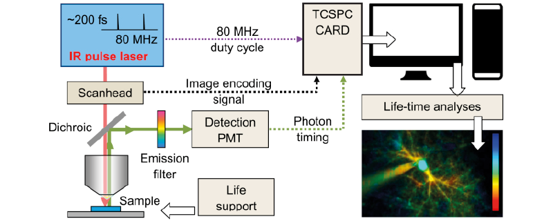
Zheng et al demonstrated in Neuron, that mapped basal Ca2+ in neurons and astrocytes with submicron resolution helps to unveil heterogeneous concentration landscapes that depend on age and preceding activity of the bain. Monitoring nanomolar-scale molecular interaction between OGB1 nanomolar sensitivity dye and Ca2+ was performed by Femto2D-Galvo equipped with FLIM module. The new generation of Femto2D-Galvo is the FEMTOSmart Galvo: read more about its technical features and benefits.